


Germ cell tumors arise from the malignant proliferation of primordial germ cells.During embryonic development, germ cells normally migrate from the yolk sac to the gonads;however, if some cells remain ectopically in the central nervous system, they can undergo malignant transformation to form tumors.
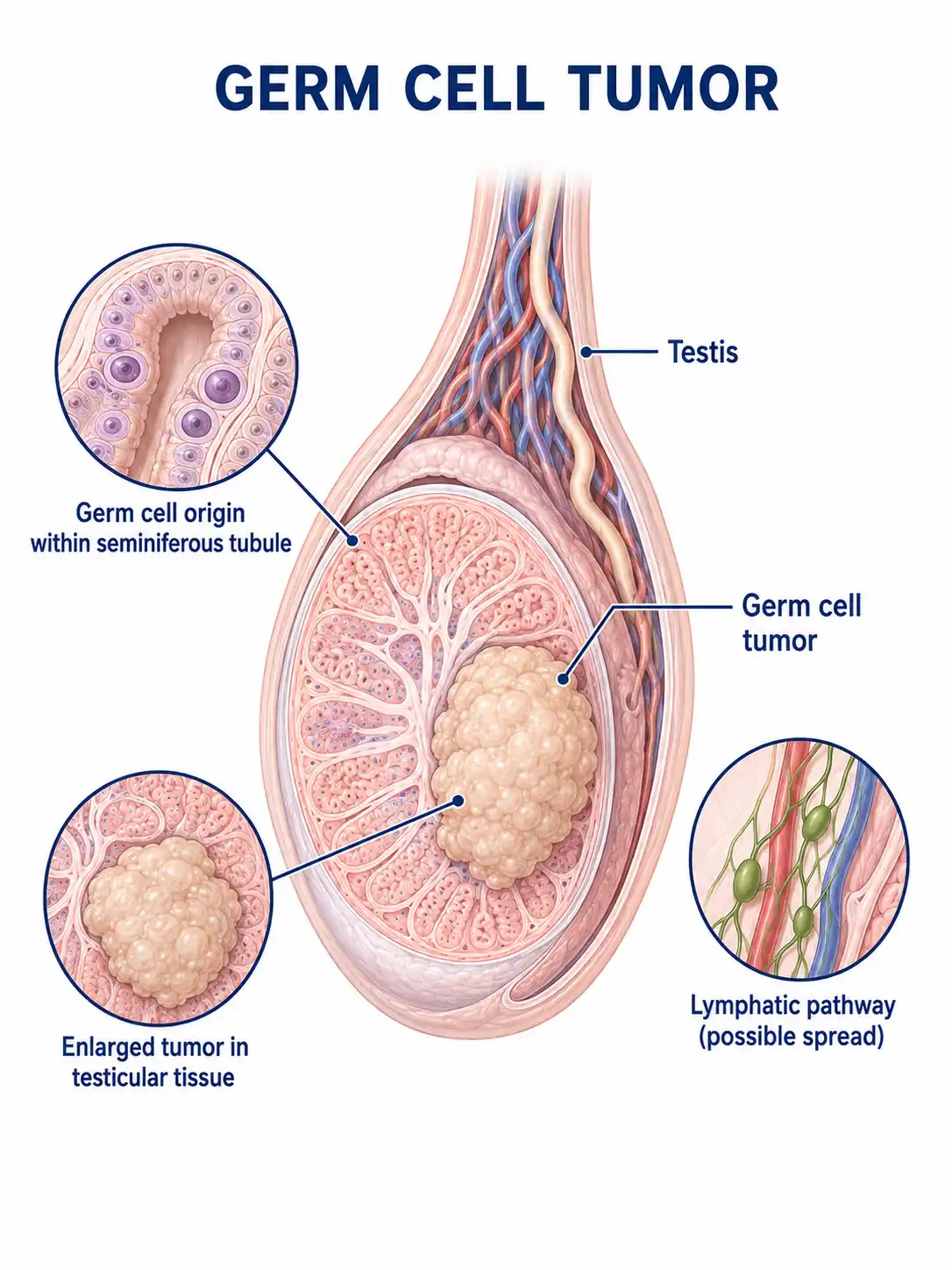
Global overview of germ cell tumors
Germ cell tumors have a relatively high incidence in Asian countries, accounting for 6%–15% of intracranial tumors in children.The pineal and suprasellar regions are the most common sites of occurrence; the condition is more common in males than in females and typically affects individuals aged 10 to 25.
Consult a doctor regarding cancer-related issues
Children or adolescents experiencing symptoms such as excessive thirst and urination, vision loss, delayed sexual development, or headache and vomiting should promptly consult a neurosurgeon or endocrinologist.
Main types of germ cell tumors
1. Germ cell tumor
Refers to pure germinoma; it is highly sensitive to radiotherapy and chemotherapy, carries the best prognosis, and may present with mildly elevated HCG levels in serum and cerebrospinal fluid.
2. Types of embryonal carcinoma
Highly malignant; classified as a non-seminomatous germ cell tumor, it shows poor sensitivity to conventional chemotherapy regimens.
3. Yolk sac tumor types
Also known as endodermal sinus tumor; secretes alpha-fetoprotein (AFP); highly malignant and rapidly progressive; requires aggressive multimodal therapy.
4. Choriocarcinoma subtype
It secretes large amounts of HCG and is highly prone to hematogenous metastasis—particularly to the lungs—making it the most malignant type of germ cell tumor.
5. Types of teratoma
Contains tissue components derived from all three germ layers; mature teratomas are benign, whereas immature teratomas and teratomas with malignant transformation possess malignant potential.
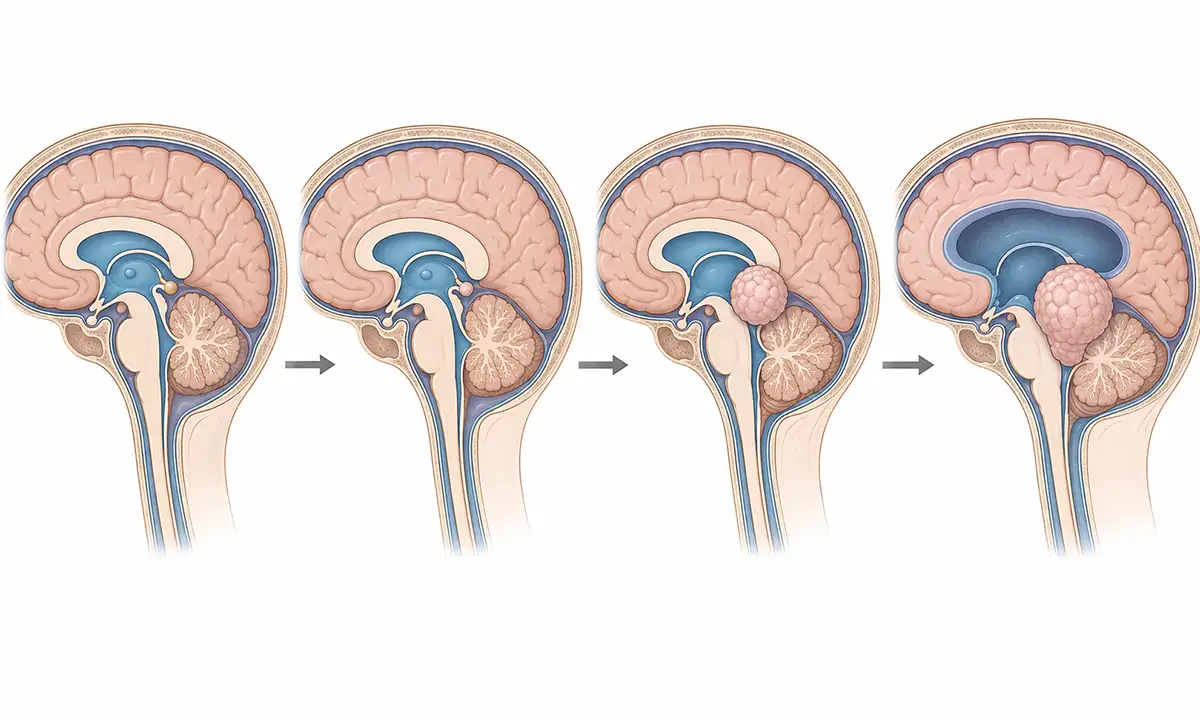
Typical symptoms and signs of germ cell tumors
1. Excessive thirst and urination
A germ cell tumor in the sellar region can compress the pituitary stalk, leading to central diabetes insipidus; symptoms include excessive thirst, polyuria, and increased nocturia, with daily urine output potentially reaching several liters.
2. Vision loss
Bitemporal hemianopia and vision loss resulting from tumor compression of the optic chiasm are the most common initial symptoms of germ cell tumors in the sellar region.
3. Abnormal sexual development
Pediatric patients may experience precocious puberty or delayed sexual development; these conditions are associated with endocrine disturbances caused by tumor-secreted hormones or compression of the pituitary gland.
4. Headache and vomiting
Pineal region tumors obstruct the cerebral aqueduct, causing obstructive hydrocephalus, which leads to headache, vomiting, and papilledema.
5. Ocular motility disorder
Tumors in the pineal region compress the superior colliculi of the quadrigeminal plate, causing Parinaud syndrome, which manifests as difficulty in upward gaze and loss of the pupillary light reflex.
6. Ataxia
Tumor compression of the cerebellum or brainstem causes unsteady gait and ataxia; severe cases may impair the ability to perform daily activities.
What are the diagnostic methods for germ cell tumors
1. Tumor marker testing
Measure serum and cerebrospinal fluid (CSF) levels of AFP and HCG; HCG may be mildly elevated in pure germinomas, AFP is elevated in yolk sac tumors, and HCG is markedly elevated in choriocarcinomas.
2. Cranial MRI scan
Contrast-enhanced MRI is the preferred method for diagnosing germinomas; these present as masses in the pineal or sellar regions with marked enhancement and the potential for cerebrospinal fluid (CSF) dissemination.
3. Cerebrospinal fluid examination
Lumbar puncture is performed to obtain cerebrospinal fluid (CSF) for cytological examination to detect shed tumor cells; a positive result confirms CSF dissemination.
4. Whole-spine MRI
Checking for spinal cord seeding lesions resulting from cerebrospinal fluid dissemination is crucial for staging and formulating the treatment plan.
5. Biopsy pathology
For tumors negative for biomarkers, stereotactic biopsy or craniotomy is required to obtain tissue samples for a definitive pathological diagnosis.
6. Endocrine assessment
Comprehensively assess pituitary function, including levels of growth hormone, thyroid hormone, cortisol, and sex hormones.
Germ Cell Tumor Treatment Methods
1. Radiotherapy
Pure germinomas are highly sensitive to radiotherapy, which serves as the primary treatment modality; this typically involves whole-ventricular irradiation followed by a boost to the tumor bed.
2. Chemotherapy
Platinum-based combination chemotherapy can eliminate micrometastases, reduce radiation dosage, and preserve cognitive function.
3. Surgical treatment
Surgical resection may be performed for residual tumor tissue following chemotherapy and radiotherapy, or in cases where the diagnosis is unclear.
4. Concurrent chemoradiotherapy
Radiotherapy combined with concurrent chemotherapy enhances efficacy and serves as the standard treatment regimen for bulky tumors or disseminated disease.
5. Cerebrospinal fluid shunting
For patients with severe obstructive hydrocephalus, perform ventriculoperitoneal shunting first to alleviate symptoms of intracranial hypertension.
6. Hormone replacement
Postoperative patients with hypopituitarism require appropriate hormone replacement therapy, including thyroxine, glucocorticoids, and sex hormones.
7. Targeted therapy
Targeted therapies for relapsed/refractory non-seminomatous germ cell tumors are currently under investigation.
8. Follow-up monitoring
Periodic follow-up with MRI and tumor marker testing after treatment is essential to monitor for recurrence and cerebrospinal fluid dissemination; long-term monitoring is crucial.
Conclusion
Germ cell tumors are malignant neoplasms originating from germ cells, frequently found in the pineal and sellar regions.With comprehensive treatment involving radiotherapy, chemotherapy, and surgery, most patients can achieve long-term survival.


