


Germ cell tumors account for 2%–3% of malignant ovarian tumors and 95% of testicular tumors. They predominantly affect children and adolescents.Pathological subtypes include teratoma, dysgerminoma, yolk sac tumor, choriocarcinoma, and embryonal carcinoma.AFP and β-hCG are important markers for diagnosis and monitoring.
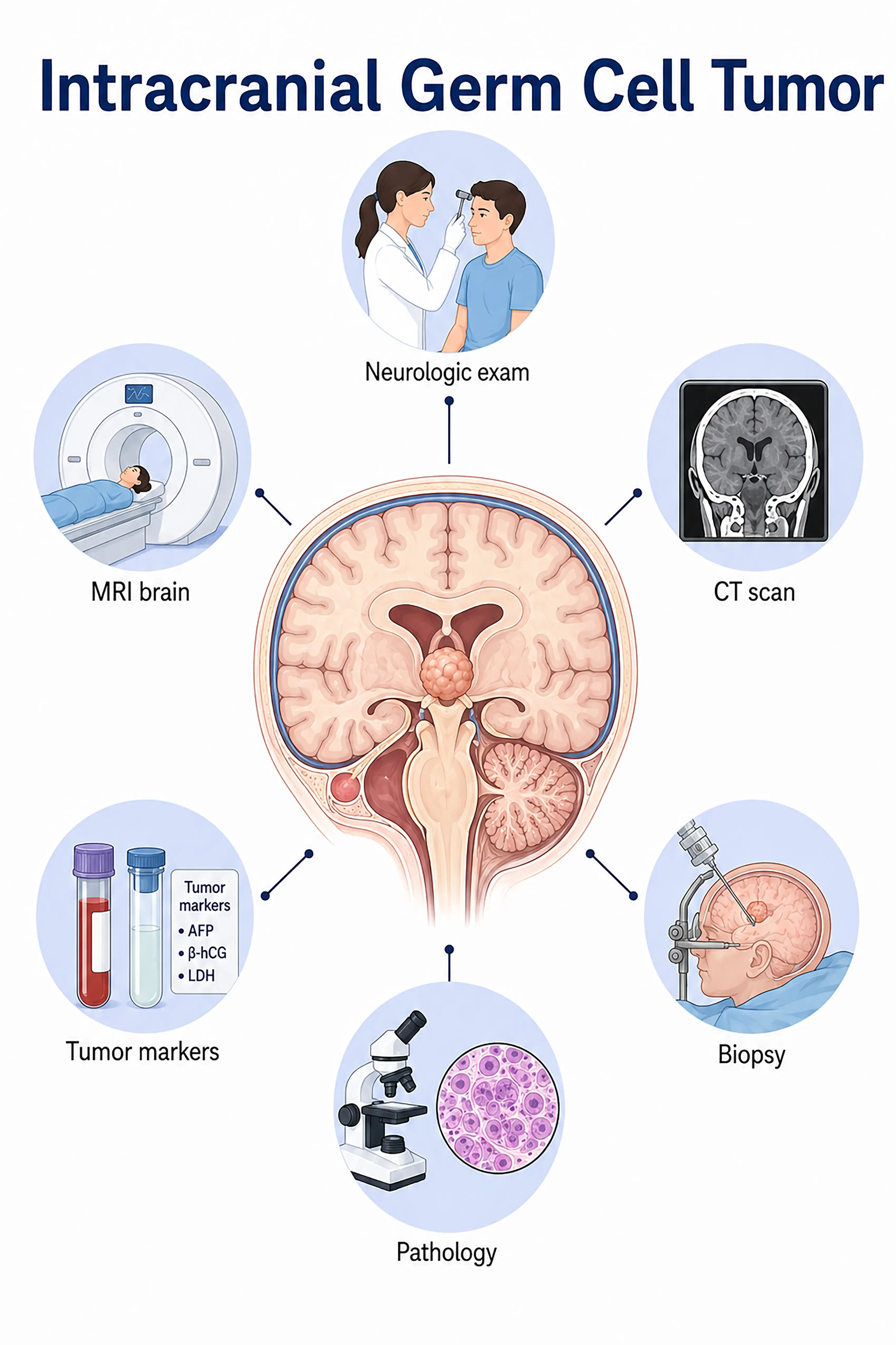
Diagnosis of germ cell tumors by stage
1.Early diagnosis of germ cell tumor
The tumor is localized; serum AFP and β-hCG levels are mildly elevated. Ultrasound or MRI shows a cystic-solid mass;abnormal tumor markers suggest a germ cell origin; diagnosis is confirmed by obtaining tissue samples during surgical exploration.
2.Intermediate-stage germ cell tumor diagnosis
Tumor enlargement accompanied by a significant rise in markers.MRI reveals tumor extent and local invasion; CT assesses lymph node metastasis; surgery or needle biopsy confirms the pathological type.
3.Advanced germ cell tumor diagnosis
Extensive tumor metastasis with significantly elevated markers.Whole-body PET-CT assesses distant metastasis; multiple biopsies confirm mixed components, guiding personalized chemotherapy regimens.
If not diagnosed promptly, malignant germ cell tumors progress rapidly and metastasize widely; however, most are sensitive to chemotherapy, and early treatment yields high cure rates.
When to Seek Medical Attention
Early signs of germ cell tumors include an abdominal mass, precocious puberty, excessive thirst and urination, and abnormally elevated serum markers.Please consult a doctor via the "Chat with a Doctor" feature to facilitate early detection.During the online consultation, the doctor will ask about your symptoms and laboratory test results, and guide you through imaging tests and a biopsy.
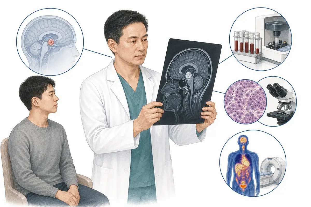
Diagnostic criteria for germ cell tumors
1.Serum AFP test
Alpha-fetoprotein (AFP) is a key marker for yolk sac tumors and embryonal carcinomas; elevated serum AFP indicates the presence of yolk sac components within the tumor and serves as a core indicator for diagnosis and monitoring treatment efficacy.
2.Serum β-hCG test
Human chorionic gonadotropin (β-hCG) levels are elevated in choriocarcinoma and some dysgerminomas; combined testing with AFP can assist in determining the pathological type of the tumor.
3.Pelvic ultrasound
Ultrasound is the preferred imaging modality; it can visualize cystic-solid masses in the ovarian or testicular regions, assess tumor size, margins, and blood flow signals, and guide needle biopsies.
4.Pelvic MRI examination
MRI offers high soft-tissue resolution, clearly demonstrating the relationship between the tumor and surrounding tissues, differentiating teratoma components (such as fat and calcification), and assessing the extent of local invasion.
5.Histopathological biopsy
Obtaining tumor tissue via surgical resection or needle biopsy for pathological examination is the gold standard for diagnosing germ cell tumors, allowing for the determination of the specific pathological type and degree of differentiation.
6.LDH level testing
Lactate dehydrogenase (LDH) levels are frequently elevated in dysgerminoma; as a marker of tumor burden, LDH can be used to monitor treatment efficacy and assess prognosis.
7.Chest CT scan
Assesses for metastases in mediastinal lymph nodes and the lungs; germ cell tumors commonly metastasize to these sites, making this a standard part of staging assessment.
8.Whole-body PET-CT
Assesses systemic metastasis, detects occult metastatic lesions that are difficult to identify via conventional imaging, and guides the formulation of chemotherapy regimens and the assessment of treatment efficacy.
9.Immunohistochemistry testing
Detection of markers such as OCT3/4, SALL4, CD117, GPC3, and PLAP assists in differentiating various types of germ cell tumors and guides precision therapy.
Daily Self-Check
Children presenting with abdominal masses, precocious puberty, or testicular enlargement in boys should seek medical attention promptly.Regular check-ups and tumor marker screening aid in early detection.
Conclusion
Diagnosis of germ cell tumors relies on tumor markers (AFP, β-hCG), imaging, and pathological examination.Most types are sensitive to chemotherapy, and early diagnosis and treatment can lead to high cure rates.


