


Gastric cancer continues to have high global incidence and mortality, but the stage at diagnosis varies markedly by region. Settings with targeted Helicobacter pylori control and endoscopic screening for high-risk groups generally detect a larger proportion of early gastric cancers, whereas advanced-stage diagnosis is more common where screening and treatment resources are limited.
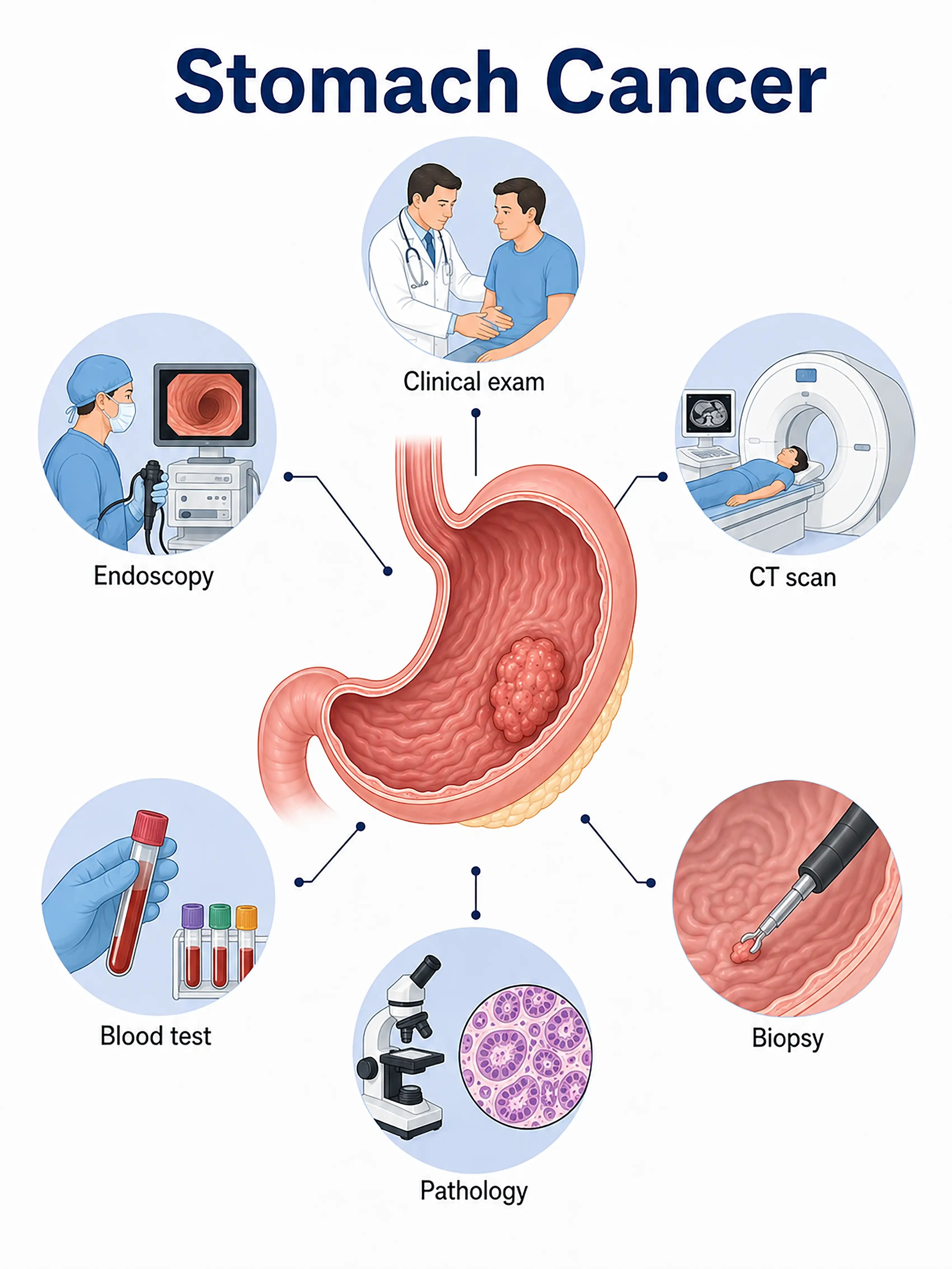
Gastric cancer diagnosis by stage
1.Early-stage gastric cancer diagnosis
The tumor is confined to the mucosa or submucosa, often without specific symptoms. Gastroscopy shows rough mucosa, erosion, or tiny ulcers. Chromoendoscopy and magnifying endoscopy aid early detection, and biopsy confirms the diagnosis.
2.Intermediate-stage gastric cancer diagnosis
The tumor invades the muscle layer or deeper. Gastroscopy shows cauliflower-like masses or ulcerative lesions. Contrast-enhanced CT assesses invasion depth and nearby nodal metastasis, while endoscopic ultrasound precisely determines T stage.
3.Advanced-stage gastric cancer diagnosis
The tumor penetrates the gastric wall and invades surrounding organs or metastasizes. Whole-body CT and PET-CT assess liver, lung, and peritoneal spread, while laparoscopy assesses peritoneal dissemination to establish stage.
Without timely diagnosis, most patients have advanced disease at diagnosis, lose the opportunity for curative surgery, and have a five-year survival rate below 50%.
When to Seek Medical Attention
Early signs include upper abdominal discomfort, reduced appetite, weight loss, and black stools. Consult a doctor through "Chat with a Doctor" for early detection. During an online consultation, the doctor will ask about your diet and symptoms and guide you toward gastroscopy.
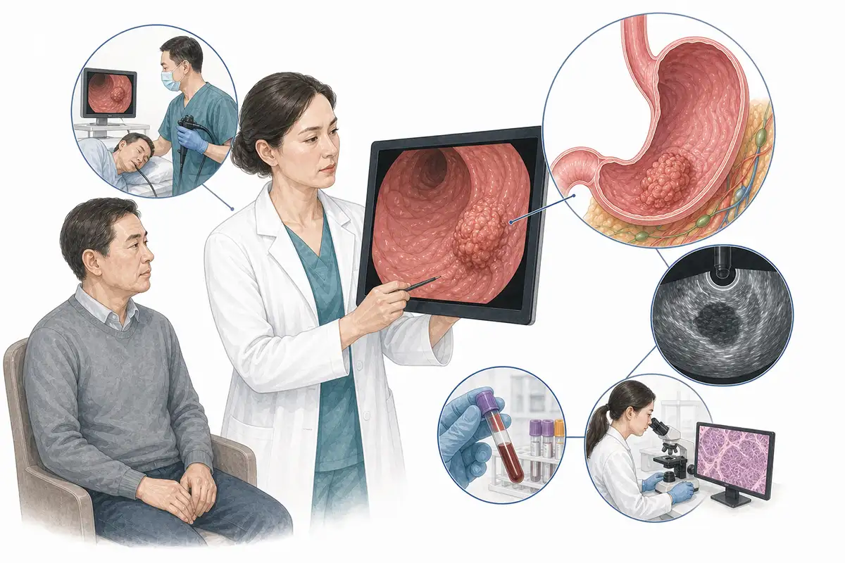
Basis for gastric cancer diagnosis
1.Gastroscopy and biopsy
Endoscopy is the first-choice diagnostic method, directly showing tumor shape, location, and extent. Forceps biopsy for pathology is the diagnostic gold standard.
2.Histopathological examination
Pathological examination of gastroscopic tissue samples determines histological type and differentiation. Gastric signet-ring cell carcinoma requires special stains for differentiation.
3.Endoscopic ultrasound
Endoscopic ultrasound clearly shows gastric wall layers and invasion depth and assesses perigastric lymph node metastasis, mainly for precise staging.
4.Abdominal CT scan
Contrast-enhanced CT is the preferred clinical staging method. Combined chest, abdominal, and pelvic scanning is recommended to assess tumor extent, nodal metastasis, and distant organ spread.
5.Barium meal X-ray examination
Double-contrast air-barium imaging is superior to conventional CT for localization, showing mucosal destruction, filling defects, and abnormal motility to support surgical selection.
6.Tumor marker testing
Serum CA19-9, CEA, CA72-4, and other tumor markers support diagnosis, response assessment, and recurrence monitoring but cannot establish a definitive diagnosis.
7.Abdominal ultrasound
This simple noninvasive examination shows gastric wall layers and surrounding lymph nodes, detects liver metastasis and ascites, and guides needle biopsy.
8.Whole-body PET-CT assessment
Whole-body PET-CT assesses distant metastasis, is recommended for suspected residual or recurrent disease, and detects lesions missed by conventional imaging.
9.Laparoscopic exploration
Assesses peritoneal metastasis difficult to detect by CT through direct abdominal exploration and tissue biopsy to establish stage.
Daily Self-Check
Pay attention to upper abdominal discomfort, reduced appetite, weight loss, and other digestive symptoms. Avoid high-salt and preserved foods, quit smoking, limit alcohol, and have regular gastroscopy if there is a family history.
Conclusion
Early gastric cancer diagnosis rates are low. Gastroscopy with pathological biopsy is the diagnostic gold standard. Greater early screening awareness is crucial to improving prognosis.


