


Gallbladder cancer is a tumor resulting from the malignant proliferation of epithelial cells in the gallbladder mucosa.The gallbladder is located in the gallbladder fossa beneath the liver; its primary functions are to store and concentrate bile, and to contract and release bile to aid digestion after meals.
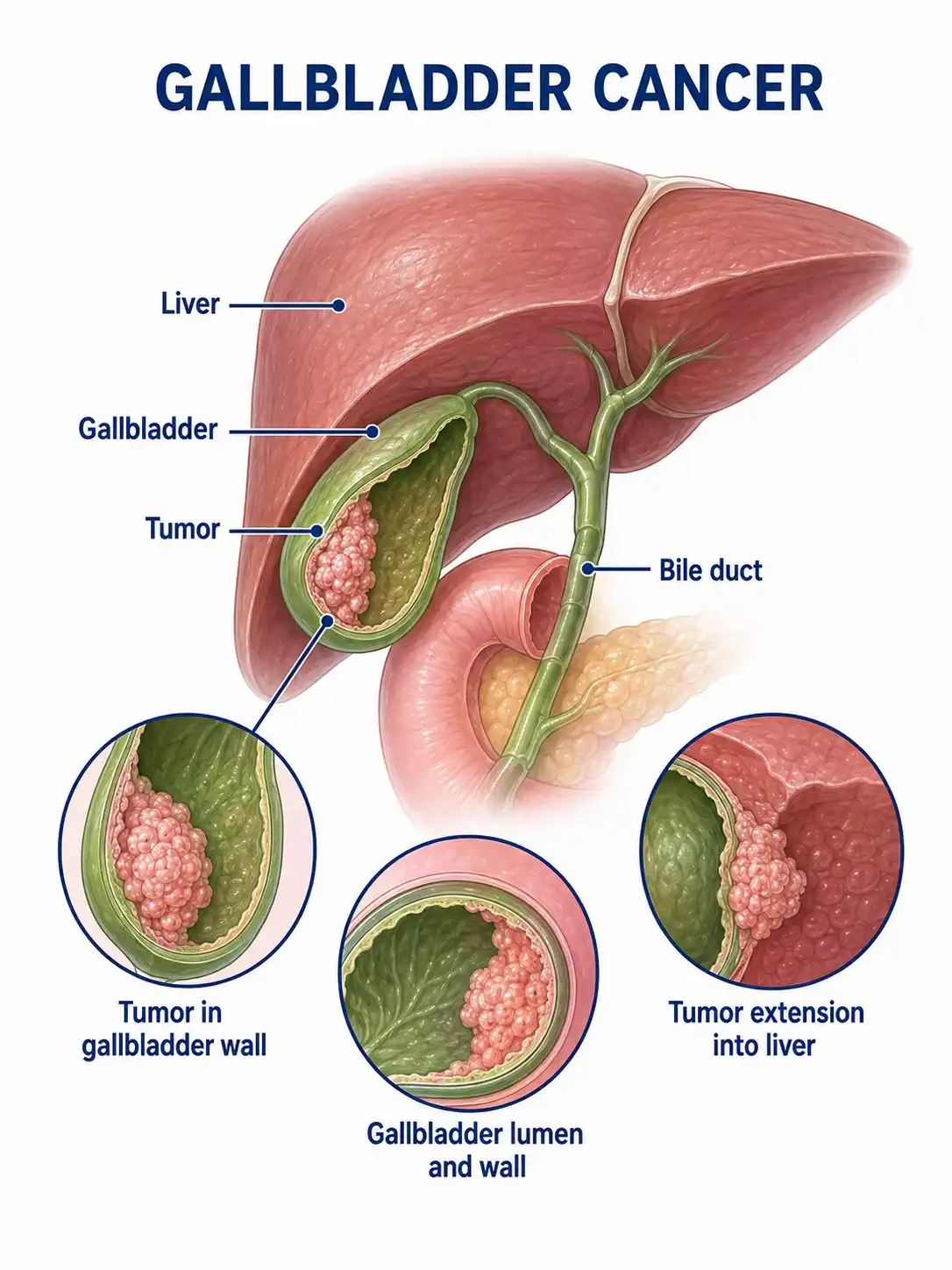
Global overview of gallbladder cancer
The incidence of gallbladder cancer varies geographically worldwide; it is more common in women than in men and is closely associated with gallstones.Incidence rates are relatively high in certain regions of my country, with the disease most frequently affecting individuals aged 60–70.
Consult a doctor regarding cancer-related issues
Individuals experiencing symptoms such as persistent right upper quadrant abdominal pain, jaundice, or a right upper quadrant abdominal mass, or those with a history of long-term gallstones, should promptly consult a hepatobiliary surgeon or a general surgeon.
Main types of gallbladder cancer
1. Types of adenocarcinoma
It is the most common type of gallbladder cancer, accounting for approximately 80%–90% of cases; subtypes include intestinal, gastric, and biliary types.
2. Squamous cell carcinoma
Relatively rare; associated with squamous metaplasia of the gallbladder mucosa; highly malignant with a poor prognosis.
3. Undifferentiated carcinoma type
Extremely rare; it is poorly differentiated, grows rapidly, and can metastasize widely at an early stage.
4. Neuroendocrine carcinoma
Originates from gallbladder endocrine cells; classified into well-differentiated neuroendocrine tumors and poorly differentiated neuroendocrine carcinomas.
5. Papillary carcinoma type
Grows in a papillary pattern into the gallbladder lumen; can cause gallbladder obstruction and acute cholecystitis.
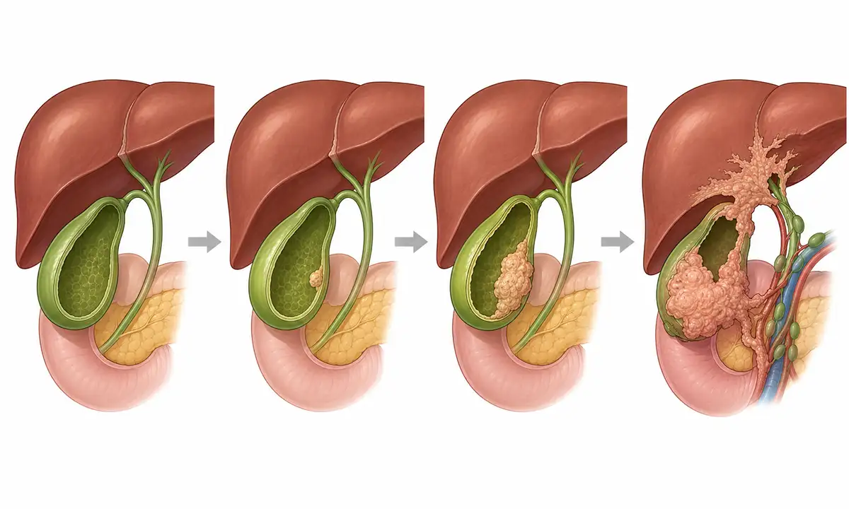
Typical symptoms and signs of gallbladder cancer
1. Right upper quadrant abdominal pain
Persistent dull pain or a sensation of fullness in the right upper abdomen is the most common symptom; it resembles chronic cholecystitis and is easily overlooked or misdiagnosed.
2. Jaundice
Obstructive jaundice occurs when the tumor invades the hilar bile ducts or metastasizes to lymph nodes in the hepatoduodenal ligament, compressing the bile ducts.
3. Right upper quadrant abdominal mass
A hard mass is palpable in the right upper quadrant when the tumor enlarges or when gallbladder hydrops develops (often associated with gallstones).
4. Weight loss
Unexplained weight loss, fatigue, and loss of appetite are significant signs of malignant tumor progression.
5. Nausea and vomiting
Presents with gastrointestinal symptoms such as loss of appetite, nausea, vomiting, and abdominal distension, associated with tumor invasion into surrounding organs.
6. Acute cholecystitis
Obstruction of the cystic duct by the tumor can trigger an acute cholecystitis attack, manifesting as severe right upper quadrant abdominal pain, fever, and a positive Murphy's sign.
What are the diagnostic methods for gallbladder cancer
1. Ultrasound examination
Abdominal ultrasound is the preferred method for screening for gallbladder cancer; it can reveal gallbladder wall thickening, masses within the gallbladder, and enlarged lymph nodes at the hepatic hilum.
2. Contrast-enhanced CT scan
Contrast-enhanced abdominal CT is the primary modality for diagnosing gallbladder cancer, allowing for the assessment of the extent of tumor invasion, liver involvement, and distant metastasis.
3. Magnetic resonance imaging (MRI)
MRI is highly valuable for the diagnosis and staging of gallbladder cancer; MRCP allows for the assessment of biliary system involvement.
4. Tumor marker
Elevated CA19-9 and CEA levels can aid in diagnosis, but specificity is low; biliary inflammation can also cause elevations.
5. Pathological biopsy
Obtaining tissue samples via surgery or needle biopsy for pathological examination is the gold standard for definitive diagnosis.
6. Chest CT scan
Chest CT is used to rule out pulmonary metastasis and is an essential component of routine preoperative staging examinations.
Gallbladder Cancer Treatment Methods
1. Cholecystectomy
Simple cholecystectomy is suitable for early-stage gallbladder cancer confined to the mucosa and submucosa.
2. Extended cholecystectomy
Resection of the gallbladder, a portion of liver tissue, regional lymph nodes, and the bile duct; suitable for cases where the tumor has invaded the full thickness of the gallbladder wall.
3. Chemotherapy
Gemcitabine combined with cisplatin can be used for postoperative adjuvant therapy or systemic treatment in patients with advanced disease to control disease progression.
4. Radiotherapy
Postoperative adjuvant radiotherapy can reduce local recurrence rates, while palliative radiotherapy can alleviate pain and symptoms of obstruction.
5. Targeted therapy
The use of drugs targeting specific molecular markers for gallbladder cancer is currently under investigation.
6. Biliary drainage
For advanced-stage patients with obstructive jaundice, biliary stent placement can improve liver function and quality of life.
7. Prophylactic resection
For high-risk individuals—such as those with gallstones accompanied by gallbladder wall calcification or polyps larger than 1 cm—prophylactic cholecystectomy may be considered.
8. Follow-up monitoring
Postoperative follow-up involving regular abdominal CT and tumor marker testing is crucial to monitor for recurrence and metastasis.
Conclusion
Gallbladder cancer is a common malignant tumor of the biliary tract and is closely associated with gallstones.Preventive surgery for high-risk populations, early detection, and standardized treatment help improve patient prognosis.


