


Gallbladder cancer is the most common malignant tumor of the biliary tract, accounting for 80%–95% of biliary tract cancers.It predominantly affects the elderly (aged 65 and older), with a higher incidence in women than in men.The risk is tenfold higher in patients with gallstones larger than 3 cm in diameter.Early-stage cases are often incidentally detected during pathological examination following a cholecystectomy.
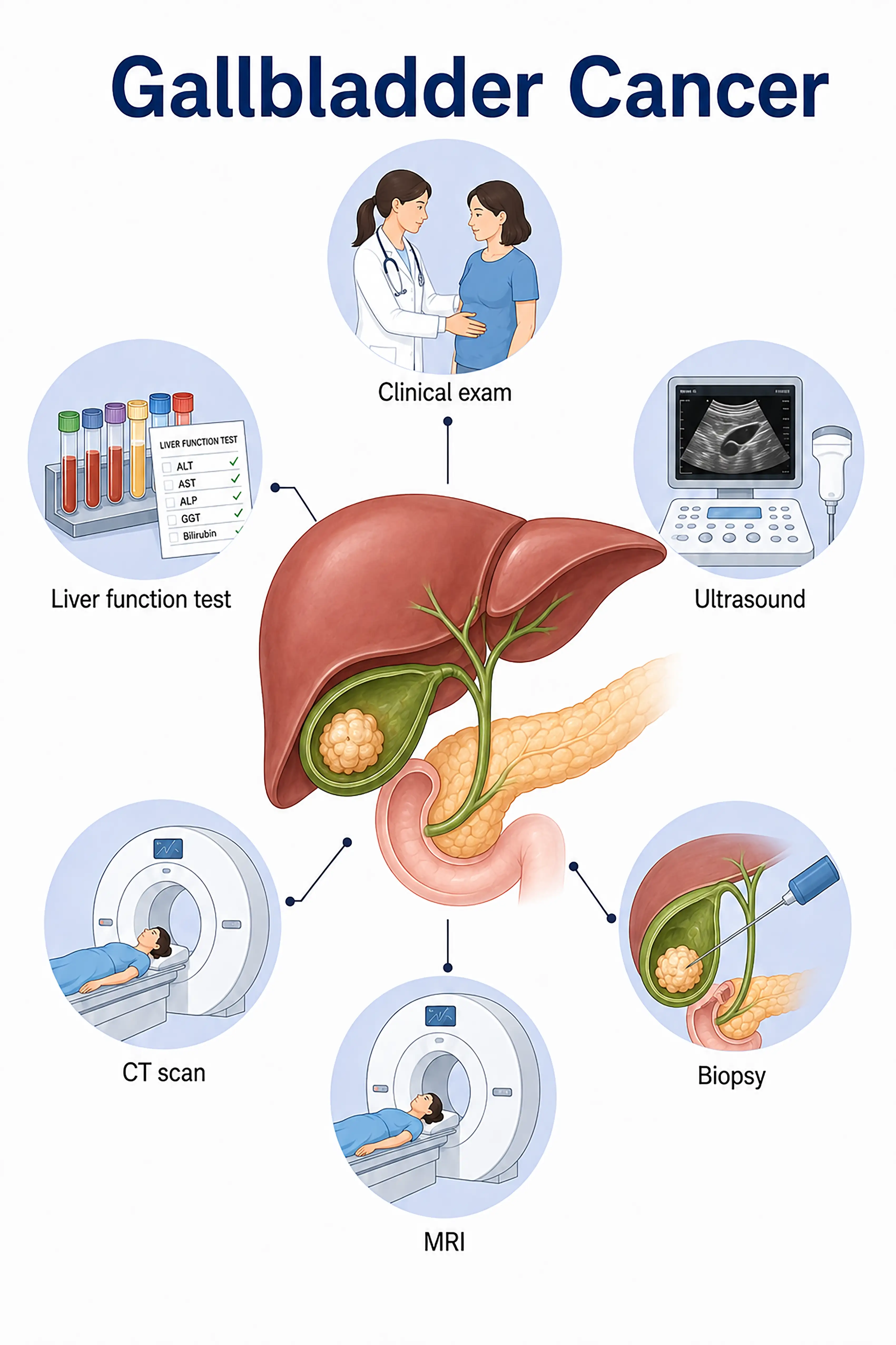
Diagnosis and staging of gallbladder cancer
1.Early diagnosis of gallbladder cancer
The tumor is confined to the gallbladder mucosa (Tis or T1a) and is often asymptomatic.Ultrasound may reveal gallbladder wall thickening or a polypoid lesion (>1 cm), followed by further evaluation via CT; diagnosis is frequently confirmed incidentally through pathological examination after cholecystectomy.
2.Diagnosis of mid-stage gallbladder cancer
Presentation includes right upper quadrant abdominal pain and jaundice.Contrast-enhanced CT and MRI show irregular gallbladder wall thickening or a mass invading the surrounding liver parenchyma; CA19-9 and CEA levels are elevated.
3.Diagnosis of advanced gallbladder cancer
Tumor extensively invades the liver and bile ducts or metastasizes distantly to the liver and peritoneum.Whole-body CT and PET-CT assess the extent of metastasis; needle biopsy confirms the pathology.
If gallbladder cancer is not diagnosed in time—as early symptoms are insidious and easily overlooked—it is often in an advanced stage by the time of diagnosis; the prognosis is extremely poor, with a five-year survival rate of less than 5%.
When to Seek Medical Attention
Early signs of gallbladder cancer include pain in the upper right abdomen, jaundice, and weight loss.Please consult a doctor via the "Chat with a Doctor" feature to facilitate early detection.During the online consultation, the doctor will ask about your history of gallbladder disease and your symptoms, and guide you through the process of getting ultrasound and CT scans.
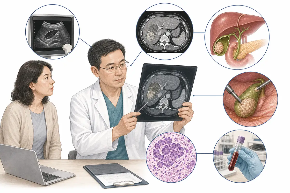
Diagnostic criteria for gallbladder cancer
1.Abdominal ultrasound
Ultrasound is the preferred method for screening for gallbladder cancer; it can visualize gallbladder wall thickening, polypoid lesions (>1 cm), or masses within the gallbladder lumen, serving as a non-invasive tool for detecting early-stage lesions.
2.Contrast-enhanced CT scan
Contrast-enhanced CT evaluates the degree of gallbladder wall thickening, mass size, and invasion of the surrounding liver parenchyma, serving as a core imaging tool for staging and the assessment of surgical resectability.
3.Abdominal MRI
MRI offers high soft-tissue resolution, enabling the assessment of the tumor's relationship with hilar vessels and bile ducts, while MRCP reveals the extent of biliary system involvement.
4.CA19-9 test
Carbohydrate antigen 19-9 (CA19-9) levels are often elevated in gallbladder cancer; combining this with CEA testing can improve diagnostic sensitivity, making it an important marker for auxiliary diagnosis and monitoring.
5.CEA testing
Carcinoembryonic antigen (CEA) levels are elevated in gallbladder cancer;combined testing with CA19-9 can improve the early detection rate.
6.Abdominal plain X-ray
Shows calcification in the gallbladder region or a porcelain gallbladder (calcification of the gallbladder wall); porcelain gallbladder is a high-risk factor for gallbladder cancer and requires surgical resection.
7.Laparoscopic exploration
Laparoscopy allows for direct visualization of the gallbladder and assessment of intra-abdominal metastasis, tumor staging, and surgical feasibility, while also enabling tissue biopsy.
8.Needle biopsy
CT-guided needle biopsy of a mass in the gallbladder region is performed to establish a pathological diagnosis and guide the treatment plan.
9.Whole-body PET-CT
Assess for distant metastasis; offers advantages in detecting occult metastatic lesions that are difficult to identify via conventional imaging, aiding in precise staging.
Daily Self-Check
Patients with gallstones should undergo regular ultrasound follow-ups; vigilance is required if significant gallbladder wall thickening or polyps larger than 1 cm are observed.Porcelain gallbladder should be surgically removed as soon as possible.
Conclusion
Early diagnosis of gallbladder cancer is challenging; ultrasound and CT scans are the primary imaging modalities.Regular follow-ups for patients with gallstones facilitate the early detection of suspicious lesions.


