


Esophageal cancer is unevenly distributed, with higher incidence in parts of Asia, the Middle East, and Africa. Risk is greater in men, and squamous cell carcinoma remains the leading type. Tobacco, alcohol, poor nutrition, very hot beverages, and chronic esophageal disease increase risk.
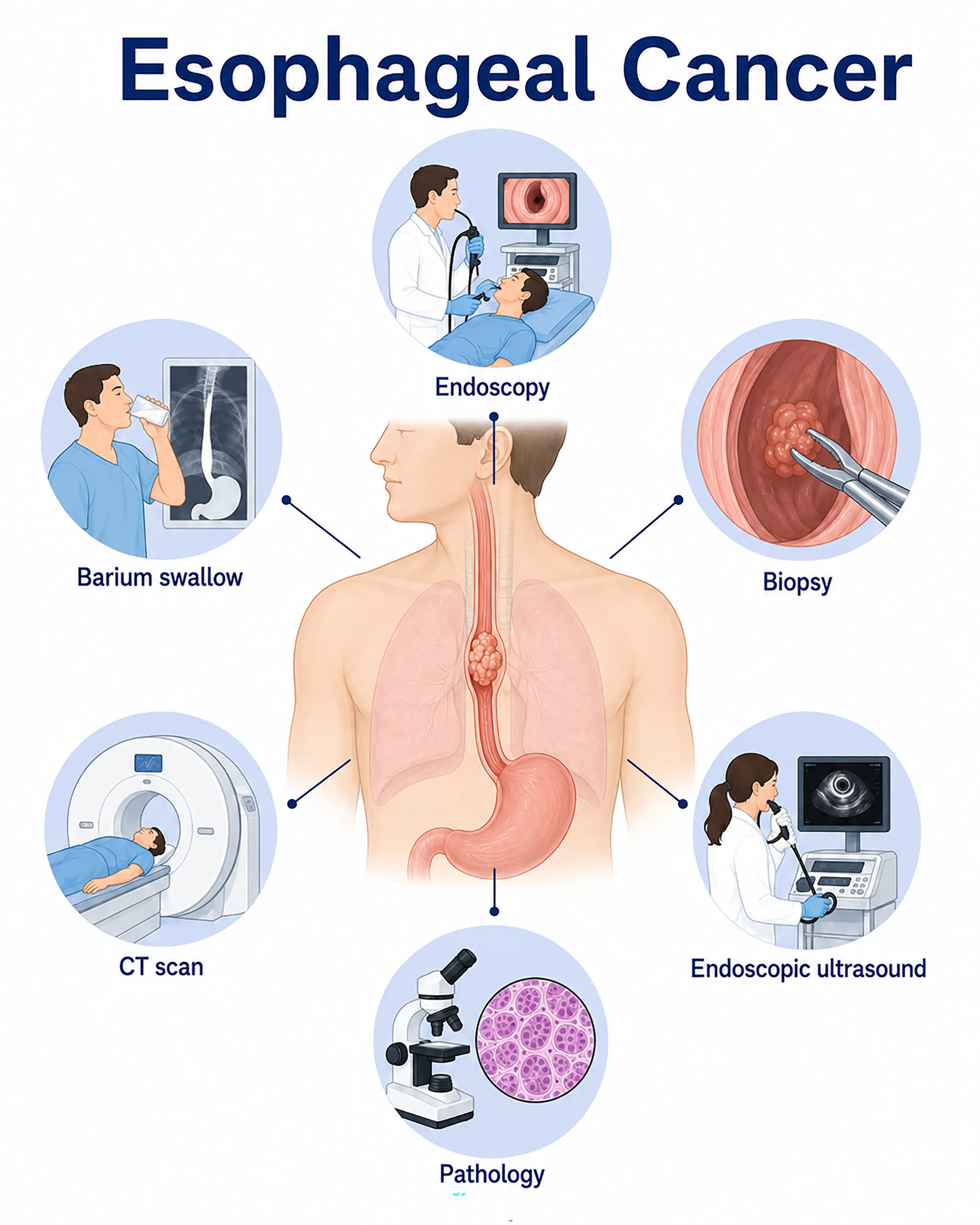
Esophageal cancer diagnosis by stage
1.Early-stage esophageal cancer diagnosis
The tumor is confined to the mucosa and symptoms are mild. Endoscopy shows rough mucosa, erosions, or small ulcers. Narrow-band imaging and chromoendoscopy detect early lesions, with biopsy confirming diagnosis.
2.Intermediate-stage esophageal cancer diagnosis
The tumor invades the muscle layer. Endoscopy shows a cauliflower-like mass or ulcerative lesion and narrowing. Contrast CT assesses invasion and nearby nodal metastases, while endoscopic ultrasound provides precise T staging.
3.Advanced-stage esophageal cancer diagnosis
The tumor penetrates the esophageal wall and invades nearby organs or spreads distantly. Whole-body CT and PET-CT assess mediastinal and abdominal nodes and liver and lung metastases. Needle biopsy establishes pathological type.
Without timely diagnosis, esophageal cancer progresses rapidly and invades nearby vital organs, eliminating the opportunity for curative surgery. Prognosis is very poor, with five-year survival below 20%.
When to Seek Medical Attention
Early warning signs include difficulty swallowing, pain behind the breastbone, food regurgitation, and unexplained weight loss. Please consult a doctor through "Chat with a Doctor" to support early detection. The doctor will ask about dietary habits and symptoms and guide you through endoscopy and upper gastrointestinal contrast imaging.
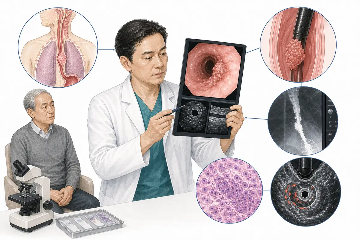
Basis for esophageal cancer diagnosis
1.Gastroscopy and biopsy
Endoscopy is the first-choice diagnostic method, directly showing tumor shape, location, and extent. Forceps biopsy for pathology is the diagnostic gold standard.
2.Barium swallow
X-rays after swallowing barium show mucosal destruction, filling defects, and narrowing, allowing assessment of esophageal motility and lesion length.
3.Endoscopic ultrasound
Endoscopic ultrasound clearly shows esophageal wall layers and invasion depth, assesses nearby nodal metastasis, and provides precise T staging.
4.Chest CT scan
Contrast-enhanced CT assesses esophageal wall invasion depth, surrounding tissue invasion, and mediastinal nodal metastases to determine resectability.
5.Upper gastrointestinal contrast imaging
Dynamic observation of barium passage shows narrowing, mucosal discontinuity, and abnormal motility, assessing esophageal patency and disease extent.
6.Abdominal CT and PET-CT
Whole-body CT and PET-CT assess distant metastases in the liver, lungs, and retroperitoneum, establishing stage and guiding combined treatment planning.
7.Narrow-band imaging endoscopy
NBI uses special wavelengths to enhance mucosal surface vessels and fine structures, helping detect early precancerous lesions and superficial cancer.
8.Chromoendoscopy
Spraying stains such as Lugol's iodine or methylene blue identifies unstained or abnormally stained lesions, improving early detection.
9.Tumor marker testing
Serum markers such as CEA, SCC, and CA199 support diagnosis and response assessment, but low specificity means they cannot establish a definitive diagnosis.
Daily Self-Check
Watch for food sticking and pain behind the breastbone, especially with dry or hard foods. Avoid prolonged consumption of very hot and pickled food, stop smoking, and limit alcohol. Those with a family history should have regular examinations and endoscopy.
Conclusion
Diagnosis relies on endoscopic biopsy and imaging-based staging. Progressive difficulty swallowing warrants prompt consultation, and early diagnosis is crucial for surgical selection.


