


Chondrosarcoma is a tumor formed by the malignant proliferation of chondrocytes.Cartilage is a vital component of the skeletal system, covering articular surfaces and forming parts of the skeleton; chondrosarcoma is characterized by the production of cartilaginous matrix by tumor cells.
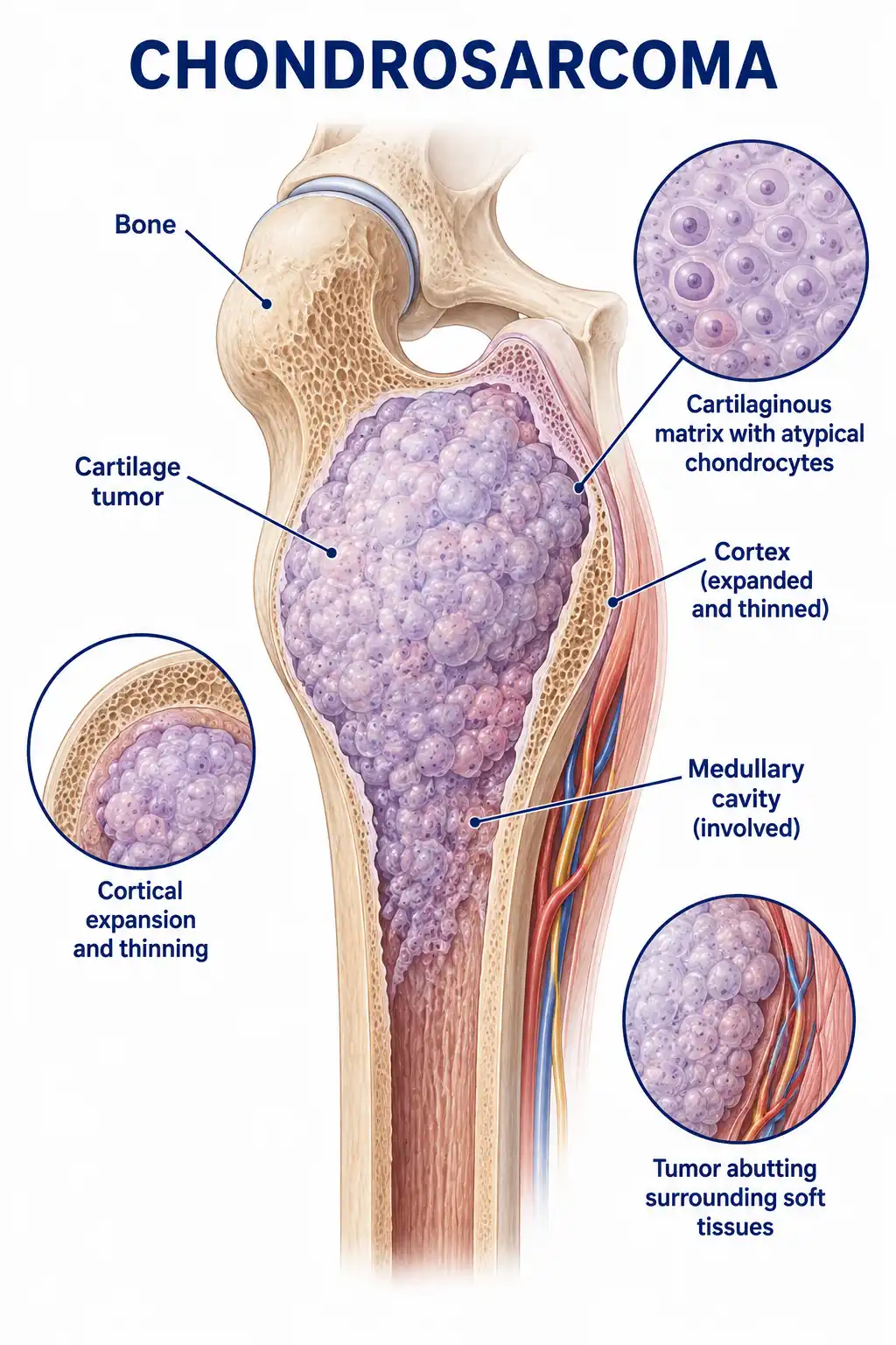
Global overview of chondrosarcoma
Chondrosarcoma is the second most common primary malignant bone tumor in adults, with an incidence of approximately 2–3 cases per million people annually.It predominantly affects middle-aged individuals (40–60 years old), with a slightly higher prevalence in men than in women; the pelvis, proximal femur, and scapula are the most common sites of occurrence.
Consult a doctor regarding cancer-related issues
Patients experiencing symptoms such as persistent bone pain, a localized mass, or the rapid enlargement of a pre-existing osteochondroma over a short period should promptly consult an orthopedic oncologist or an orthopedist.
Main types of chondrosarcoma
1. Conventional chondrosarcoma
The most common type, accounting for approximately 85% of chondrosarcomas; it typically occurs in the pelvis and proximal femur and is classified into grades I–III, with higher grades indicating a poorer prognosis.
2. Mesenchymal chondrosarcoma
A highly malignant subtype composed of undifferentiated small round cells and well-differentiated cartilage islands; it commonly occurs in the jawbones and ribs.
3. Dedifferentiated chondrosarcoma
Low-grade chondrosarcoma with a high-grade non-chondrosarcomatous sarcomatous component carries an extremely poor prognosis and a high risk of distant metastasis.
4. Clear cell chondrosarcoma
A low-grade malignant tumor that typically occurs in the proximal femoral epiphysis; it has a relatively good prognosis but is prone to local recurrence.
5. Secondary chondrosarcoma
Arises from the malignant transformation of pre-existing benign cartilaginous lesions, such as osteochondromas or enchondromas; it accounts for approximately 10% of chondrosarcomas.
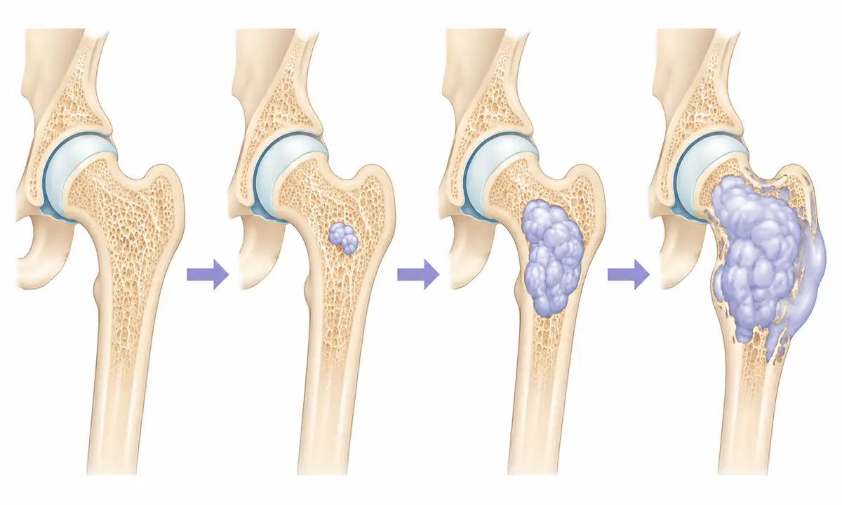
Typical symptoms and signs of chondrosarcoma
1. Bone pain
Progressive bone pain is the most common symptom; it begins as a dull ache or soreness, intensifies as the tumor grows, and is notably worse at night.
2. Localized mass
Tumor growth results in a hard, fixed mass at the affected site; the mass has a smooth surface, grows slowly, and may have a history spanning months or even years.
3. Joint dysfunction
When the tumor is located near a joint, it can cause restricted joint movement and pain; pelvic tumors can cause sciatica.
4. Compression symptoms
Spinal and pelvic tumors can compress the spinal cord or nerve roots, causing limb numbness, weakness, and even bowel or bladder dysfunction.
5. Pathologic fracture
Tumor destruction of the bone cortex reduces bone strength, potentially leading to pathological fractures.
6. Systemic symptoms
Patients in the advanced stage exhibit systemic symptoms such as weight loss and fatigue; the dedifferentiated type is prone to pulmonary metastasis.
Diagnostic methods for chondrosarcoma
1. X-ray examination
X-ray imaging reveals bone destruction accompanied by punctate or annular calcification, as well as expansion and thinning or thickening of the cortex, aiding in preliminary diagnosis.
2. CT scan
CT can clearly demonstrate intratumoral calcification patterns and cortical bone destruction, facilitating the assessment of the relationship between the tumor and surrounding tissues.
3. Magnetic resonance imaging (MRI)
MRI is the best method for assessing the intramedullary and soft-tissue extent of a tumor, aiding in surgical planning.
4. Bone scan
Whole-body bone scanning allows for the assessment of multiple lesions and bone metastases, providing a valuable reference for staging.
5. Biopsy pathology
Pathological examination of tumor tissue obtained via needle or incisional biopsy is the gold standard for definitive diagnosis and allows for the determination of the tumor grade.
6. Molecular testing
Detecting molecular markers such as IDH1/2 mutations aids in differential diagnosis and prognostic assessment.
Treatment Methods for Chondrosarcoma
1. Wide resection
Wide resection is the primary treatment for chondrosarcoma; ensuring adequate safety margins is crucial for preventing recurrence.
2. Reconstructive surgery
Following tumor resection, limb function is reconstructed via artificial joint replacement, allogeneic bone grafting, or autologous bone grafting.
3. Pelvic surgery
Surgery for pelvic chondrosarcoma is complex;it may require hemipelvectomy and reconstruction, and functional recovery post-surgery necessitates long-term rehabilitation.
4. Radiotherapy
Chondrosarcoma has low sensitivity to radiotherapy;radiation is primarily used as an adjuvant therapy for tumors that cannot be completely resected or for recurrent tumors.
5. Chemotherapy
Conventional chondrosarcoma is insensitive to chemotherapy, whereas chemotherapy may be considered for mesenchymal and dedifferentiated subtypes.
6. Targeted therapy
Targeted drugs for IDH1 mutations and mTOR inhibitors are being studied for advanced chondrosarcoma.
7. Spinal surgery
Spinal chondrosarcoma requires surgical resection and reconstruction of spinal stability;the surgery is complex, and adjuvant radiotherapy is often required postoperatively.
8. Follow-up monitoring
Postoperative follow-up involving regular imaging is crucial to monitor for local recurrence and pulmonary metastasis.
Conclusion
Chondrosarcoma is a common primary malignant bone tumor in adults, with extensive surgical resection serving as the primary treatment.Early diagnosis and standardized treatment help improve patient prognosis.


