


Chondrosarcoma is the second most common primary malignant bone tumor, accounting for approximately 10%–20% of all bone tumors.It typically affects adults aged 40–60, with a higher incidence in men than in women.It most commonly occurs in the pelvis, femur, and scapula.Low-grade tumors have a better prognosis, whereas high-grade tumors are prone to recurrence and metastasis.
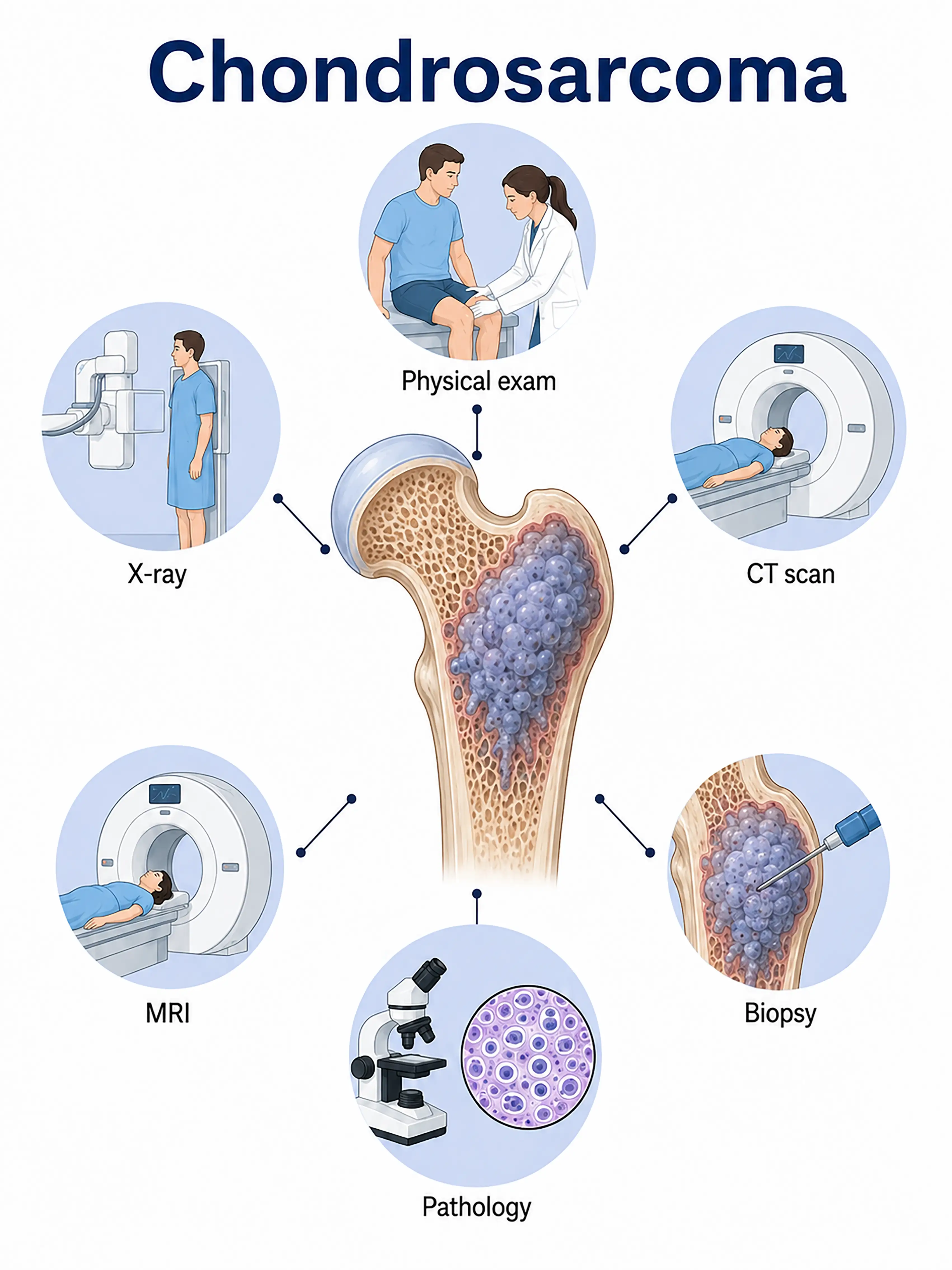
Diagnosis by stage of chondrosarcoma
1.Early diagnosis of chondrosarcoma
The tumor is confined to the bone, grows slowly, and presents with intermittent pain.X-rays show osteolytic destruction with characteristic chondroid calcification (ring-like or arc-like); further evaluation is performed via CT and MRI, and the diagnosis is confirmed by needle biopsy.
2.Diagnosis of intermediate-stage chondrosarcoma
Pain intensifies, and a localized mass appears.CT scans reveal calcification and cortical bone destruction; MRI assesses the extent of soft-tissue invasion, and biopsy determines the pathological grade.
3.Diagnosis of advanced chondrosarcoma
The tumor extensively invades surrounding tissues or metastasizes to the lungs.Whole-body CT and PET-CT are used to assess the extent of metastasis, and surgical resection provides a complete specimen to determine the grade.
If chondrosarcoma is not diagnosed promptly, high-grade tumors will invade surrounding tissues and metastasize to distant sites; the local recurrence rate is high, and the prognosis worsens significantly.
When to Seek Medical Attention
Early signs of chondrosarcoma include persistent bone pain, enlargement of a localized mass, and restricted movement.Please consult a doctor via the "Chat with Doctor" feature to facilitate early detection.During the online consultation, the doctor will ask about the characteristics of your pain and any changes in the mass, and guide you regarding imaging tests and biopsies.
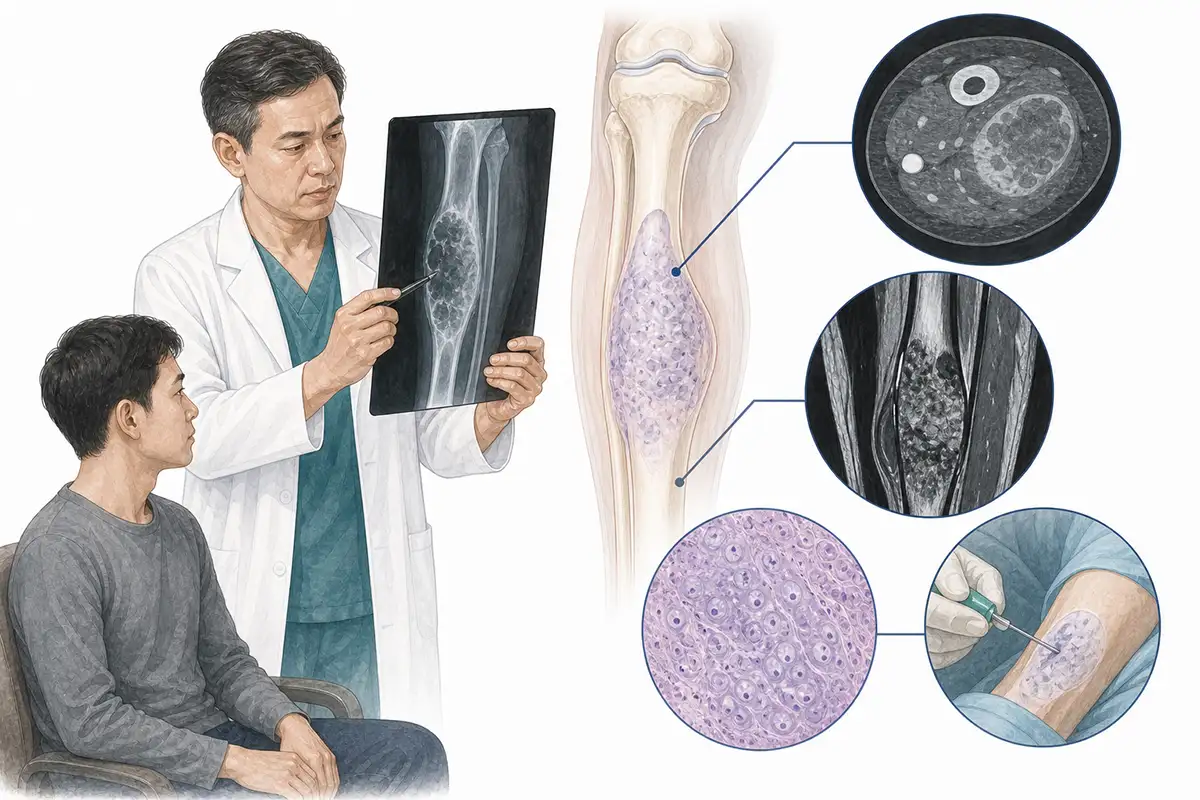
Basis for diagnosing chondrosarcoma
1.Plain X-ray examination
X-ray imaging serves as the initial imaging modality for chondrosarcoma; typical findings include osteolytic destruction accompanied by characteristic chondroid calcification (ring-like, arc-like, or popcorn-like patterns).
2.CT scan
CT clearly displays the distribution and characteristics of chondroid calcification, cortical bone destruction, and soft-tissue masses, serving as a vital tool for assessing tumor extent and formulating surgical plans.
3.MRI examination
MRI reveals long T1 and long T2 signals in the non-calcified portions of the tumor, enabling the assessment of the extent of intramedullary infiltration and involvement of surrounding soft tissues.
4.Needle biopsy
CT-guided needle biopsy for pathological examination is the gold standard for diagnosing chondrosarcoma; differential diagnosis from lesions such as enchondroma is required.
5.Whole-body bone scan
Radionuclide bone scanning shows tracer uptake at the lesion site, ruling out multifocal bone lesions and distant bone metastases.
6.Chest CT scan
Assesses for pulmonary metastases; high-grade chondrosarcomas are prone to pulmonary metastasis, making this a standard examination for initial staging.
7.PET-CT assessment
Whole-body PET-CT assesses distant metastasis and local recurrence; high-grade chondrosarcomas exhibit high metabolic activity, aiding in the differentiation between benign and malignant lesions.
8.Differential diagnosis assessment
Differential diagnosis is required to distinguish it from conditions such as enchondroma, chondroblastoma, osteosarcoma, and chordoma;assessment should integrate imaging and pathological characteristics.
9.Postoperative pathological grading
Pathological examination of the surgical specimen determines the grade according to the WHO grading system (Grades I–III), guiding postoperative follow-up and treatment plans.
Daily Self-Check
Persistent bone pain or a mass in the bone region warrants timely medical attention.Individuals with a family history of bone tumors or a history of bone disease should undergo regular imaging follow-ups.
Conclusion
Diagnosis of chondrosarcoma relies on X-ray, CT, MRI, and pathological biopsy. It requires differentiation from various benign bone tumors;early, complete surgical resection is the optimal treatment.


