


Cholangiocarcinoma is a tumor arising from the malignant proliferation of bile duct epithelial cells.The bile ducts form the system that transports bile; they are categorized into intrahepatic and extrahepatic bile ducts, with the latter further divided into hilar and distal bile ducts.
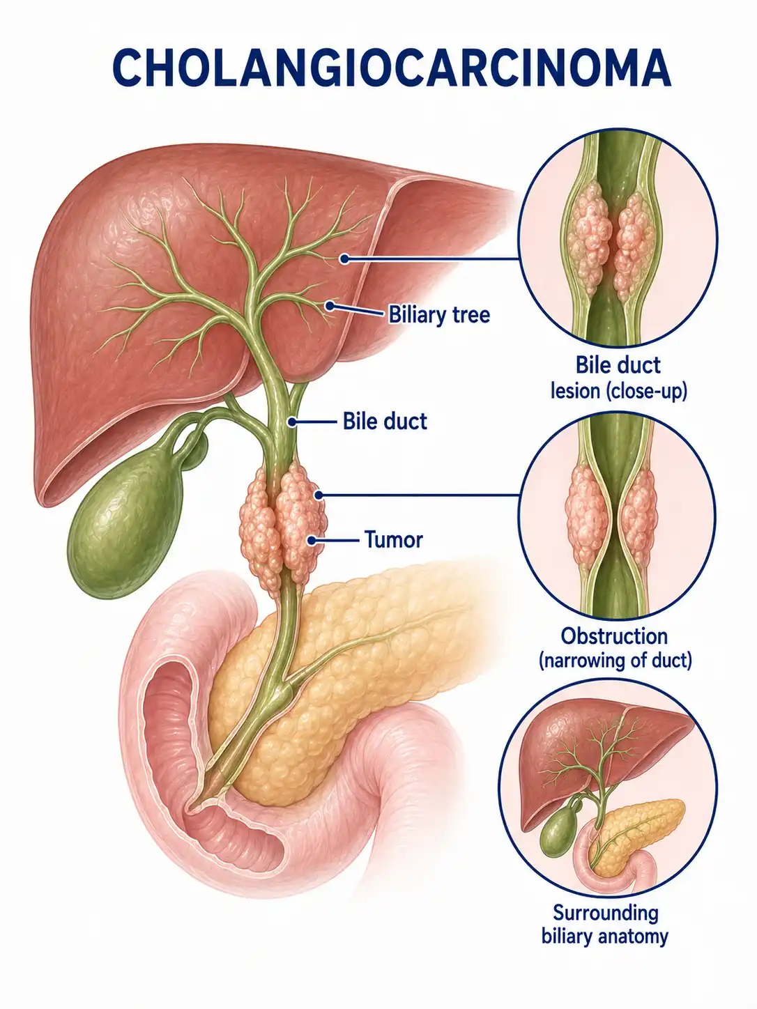
Global overview of cholangiocarcinoma
The incidence of cholangiocarcinoma varies geographically; rates are higher in Southeast Asia, linked to liver fluke infections.In my country, the incidence is rising, with the disease most commonly affecting individuals aged 50–70.
Consult a doctor regarding cancer-related issues
Individuals presenting with symptoms such as painless progressive jaundice, pruritus (itching), clay-colored stools, or right upper quadrant abdominal pain should promptly consult a hepatobiliary surgeon or gastroenterologist.
Main types of cholangiocarcinoma
1. Intrahepatic cholangiocarcinoma
Originates from intrahepatic bile ducts proximal to the secondary branches; histologically an adenocarcinoma; incidence is relatively low but has shown an upward trend in recent years.
2. Hilar cholangiocarcinoma
Also known as Klatskin tumor; arises at the junction of the left and right hepatic ducts; the most common type of extrahepatic cholangiocarcinoma; surgically challenging.
3. Distal bile duct carcinoma
Occurs in the pancreatic segment and the intraduodenal segment of the common bile duct; differential diagnosis is required to distinguish it from carcinoma of the head of the pancreas and ampullary carcinoma.
4. Papillary carcinoma
It grows in a papillary pattern into the lumen, causing bile duct dilation and obstructive jaundice, and carries a relatively favorable prognosis.
5. Sclerosing carcinoma
Infiltrative growth along the bile duct wall causes wall thickening and luminal narrowing; early diagnosis is difficult, and the prognosis is poor.
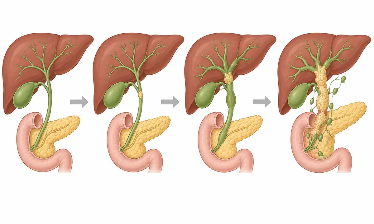
Typical symptoms and signs of cholangiocarcinoma
1. Painless jaundice
Progressively worsening painless obstructive jaundice is the most typical symptom, manifesting as yellowing of the skin and sclera, dark urine, and clay-colored stools.
2. Skin itching
Deposition of bile salts in the skin causes intractable pruritus that worsens at night, severely affecting sleep and quality of life.
3. Right upper quadrant abdominal pain
Persistent dull pain or colic in the right upper quadrant occurs due to tumor enlargement or associated cholangitis; the pain may radiate to the right shoulder and back.
4. Weight loss
Symptoms such as unexplained weight loss, fatigue, and loss of appetite are associated with impaired digestion and absorption caused by biliary obstruction.
5. Fever and chills
The presence of Charcot's triad—high fever, chills, and abdominal pain—in the context of a concurrent biliary tract infection is a classic manifestation of acute cholangitis.
6. Gallbladder enlargement
Distal bile duct carcinoma causes cholestatic enlargement of the gallbladder, presenting as a palpable, smooth, elastic, and painless gallbladder.
Diagnostic Methods for Cholangiocarcinoma
1. Liver function test
Significant elevations in serum total bilirubin and direct bilirubin, along with elevated alkaline phosphatase and gamma-glutamyl transferase, indicate obstructive jaundice.
2. Contrast-enhanced CT scan
Contrast-enhanced abdominal CT can reveal bile duct dilation, wall thickening, and masses, while assessing vascular invasion and lymph node metastasis.
3. Magnetic resonance cholangiopancreatography (MRCP)
MRCP enables 3D reconstruction of the biliary system and clearly visualizes the location, extent, and severity of bile duct strictures; it is the gold standard for non-invasive examination.
4. Tumor marker
CA19-9 is the most commonly used serum marker for cholangiocarcinoma; significant elevation suggests a possible malignancy, though biliary inflammation can also cause elevations.
5. Endoscopic retrograde cholangiopancreatography (ERCP)
ERCP allows for direct visualization of bile duct lesions and tissue biopsy, while also enabling biliary stent placement for drainage and jaundice relief.
6. Percutaneous transhepatic cholangiography
PTC is indicated for bile duct obstruction located at or above the hepatic hilum; it visualizes the bile ducts proximal to the obstruction and guides percutaneous drainage procedures.
Cholangiocarcinoma Treatment Methods
1. Surgical resection
Radical surgery is the only means of achieving long-term survival for cholangiocarcinoma; the procedure involves liver resection combined with bile duct resection and lymph node dissection.
2. Liver transplantation
Liver transplantation may be considered for strictly selected patients with hilar cholangiocarcinoma; combining it with neoadjuvant chemoradiotherapy can improve therapeutic outcomes.
3. Biliary drainage
Biliary stent placement via ERCP or PTCD can alleviate obstructive jaundice and improve liver function, creating favorable conditions for subsequent treatment.
4. Chemotherapy
Gemcitabine combined with cisplatin is a first-line chemotherapy regimen for advanced cholangiocarcinoma; it can control disease progression and prolong survival.
5. Radiotherapy
Postoperative adjuvant radiotherapy can reduce local recurrence rates, and stereotactic radiotherapy can be used for unresectable locally advanced tumors.
6. Targeted therapy
Drugs targeting FGFR2 fusions, IDH1 mutations, and other targets provide new treatment options for advanced cholangiocarcinoma.
7.Immunotherapy
PD-1 inhibitor therapy may be considered for MSI-H/dMMR cholangiocarcinoma; while the patient population that benefits is limited, the therapeutic effect is durable.
8. Palliative support
For patients with advanced disease, provide comprehensive treatment aimed at controlling jaundice, relieving pain, and improving nutritional status.
Conclusion
Cholangiocarcinoma is a malignant tumor originating from the bile duct epithelium; early symptoms are often inconspicuous.Prognosis can be improved through increased awareness, seeking timely medical attention upon the onset of painless jaundice, and undergoing comprehensive treatment centered on standardized surgery.


