


Cholangiocarcinoma is a relatively rare malignant tumor, accounting for approximately 3% of all gastrointestinal malignancies.It is classified by location into intrahepatic cholangiocarcinoma and extrahepatic cholangiocarcinoma (including hilar cholangiocarcinoma and distal cholangiocarcinoma).Primary sclerosing cholangitis and liver fluke infection are major risk factors.
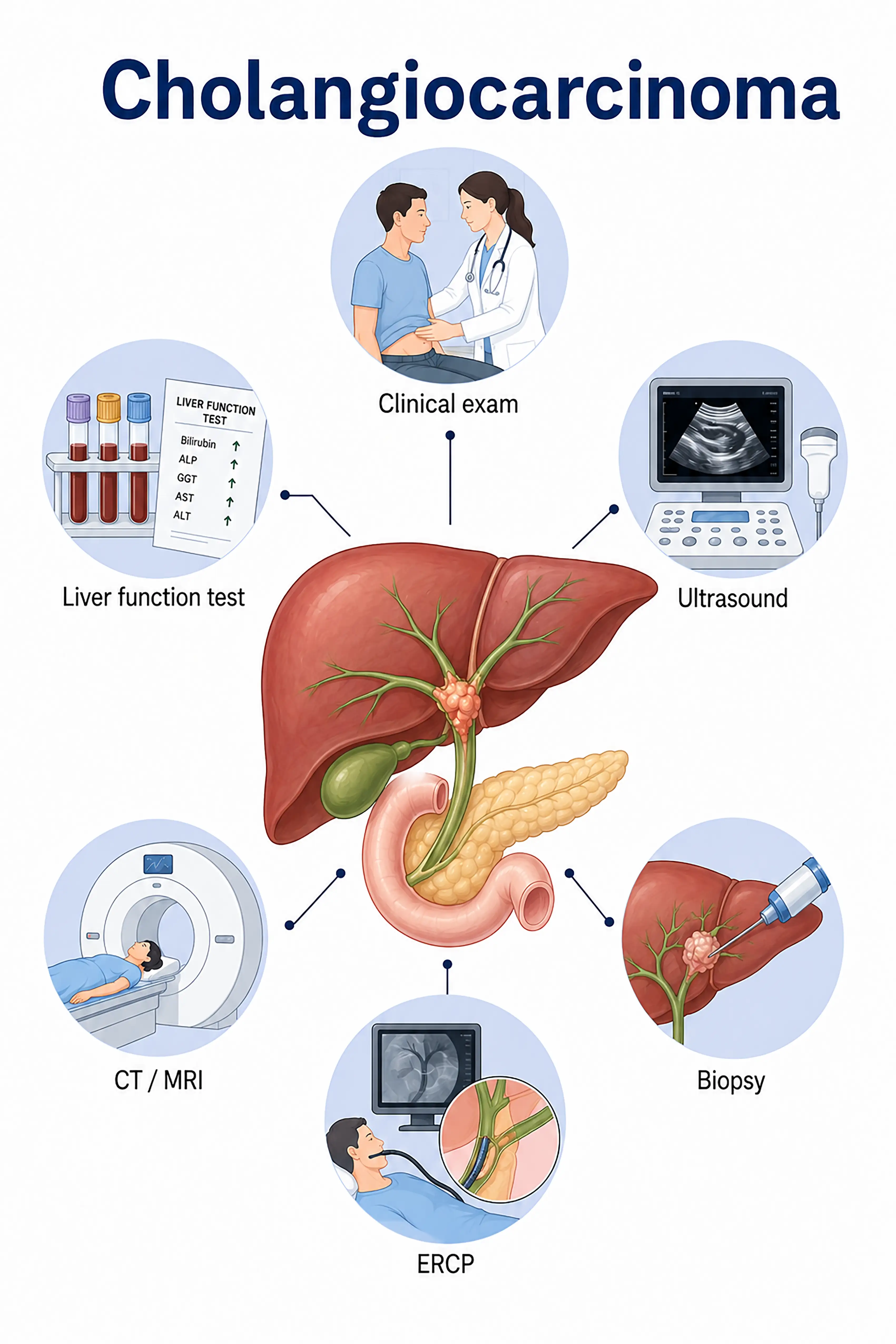
Diagnosis based on cholangiocarcinoma stages
1.Early diagnosis of bile duct cancer
The tumor is confined to the mucosal layer of the bile duct and typically presents as painless jaundice.MRCP reveals localized bile duct stenosis or a filling defect; CA19-9 levels are mildly elevated; diagnosis is confirmed via ERCP-guided brushing or biopsy.
2.Diagnosis of mid-stage bile duct cancer
The tumor invades the full thickness of the bile duct wall or surrounding tissues.Contrast-enhanced CT and MRI reveal bile duct wall thickening and surrounding infiltration; ERCP or PTC is used to obtain tissue samples for pathological confirmation.
3.Diagnosis of advanced bile duct cancer
Tumor extensively invades the hepatic parenchyma or metastasizes distantly to the liver and peritoneum.Whole-body CT and PET-CT assess the extent of metastasis; needle biopsy confirms the pathological type.
If not diagnosed promptly, cholangiocarcinoma leads to persistent biliary obstruction, liver failure, and unresectable extensive metastasis, resulting in a very poor prognosis.
When to Seek Medical Attention
Early signs of cholangiocarcinoma include jaundice, itchy skin, clay-colored stools, and pain in the upper right abdomen.Please consult a doctor via the "Chat with Doctor" feature for early detection.During the online consultation, the doctor will ask about your symptoms and medical history, and guide you through MRCP and liver function tests.
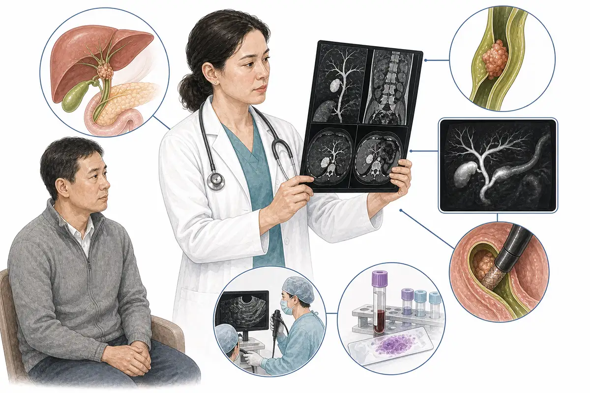
Basis for Diagnosing Cholangiocarcinoma
1.MRCP examination
MRCP is the preferred non-invasive method for diagnosing cholangiocarcinoma; it clearly visualizes the entire biliary tree, localizes sites of stenosis or obstruction, and assesses the extent of the tumor.
2.Contrast-enhanced CT scan
Contrast-enhanced CT evaluates bile duct wall thickening, liver parenchyma invasion, and lymph node metastasis, demonstrating the Bismuth-Corlette classification of hilar cholangiocarcinoma.
3.CA19-9 test
Carbohydrate antigen 19-9 (CA19-9) is the most important serum marker for cholangiocarcinoma; elevated levels suggest a possible diagnosis of cholangiocarcinoma, though cholangitis can also cause elevations, necessitating differential diagnosis.
4.ERCP examination
Endoscopic retrograde cholangiopancreatography (ERCP) allows for direct visualization of bile duct lesions and the collection of cytologic specimens via brushing or biopsy, while also facilitating stent placement for drainage.
5.Percutaneous transhepatic cholangiography
Percutaneous transhepatic cholangiography (PTC)—involving the injection of contrast medium into the intrahepatic bile ducts—visualizes the biliary system; it is suitable for patients with hilar obstruction and allows for bile collection for cytologic examination.
6.Endoscopic ultrasound
EUS assesses the layered structure of the bile duct wall and regional lymph nodes; ultrasound-guided fine-needle aspiration (FNA) of lymph nodes yields cytologic specimens, aiding in staging and diagnosis.
7.Liver function test
Measures levels of total bilirubin, direct bilirubin, ALP, and GGT to assess the severity of biliary obstruction and the extent of liver function impairment.
8.Biliary tract biopsy/brushing
Collecting bile duct epithelial cells via a cytology brush or obtaining tissue samples via forceps biopsy during ERCP are key steps for definitive diagnosis.
9.Whole-body PET-CT
Assess for distant metastasis; offers advantages in detecting occult metastatic lesions that are difficult to identify via conventional imaging, aiding in precise staging.
Daily Self-Check
Seek medical attention promptly if unexplained jaundice, pruritus, or clay-colored stools occur.Patients with a history of primary sclerosing cholangitis should undergo regular monitoring of CA19-9 levels and MRCP.
Conclusion
Diagnosis of cholangiocarcinoma relies on MRCP, ERCP, and CA19-9 testing.Early detection and timely relief of biliary obstruction are crucial for improving prognosis.


