


Cervical cancer prevalence worldwide
Cervical cancer is one of the most common malignancies among women worldwide, with a particularly high incidence and mortality burden in low- and middle-income countries and regions. Persistent infection with high-risk HPV is the main cause. Expanding HPV vaccination, screening coverage, and early treatment can effectively reduce both incidence and mortality.
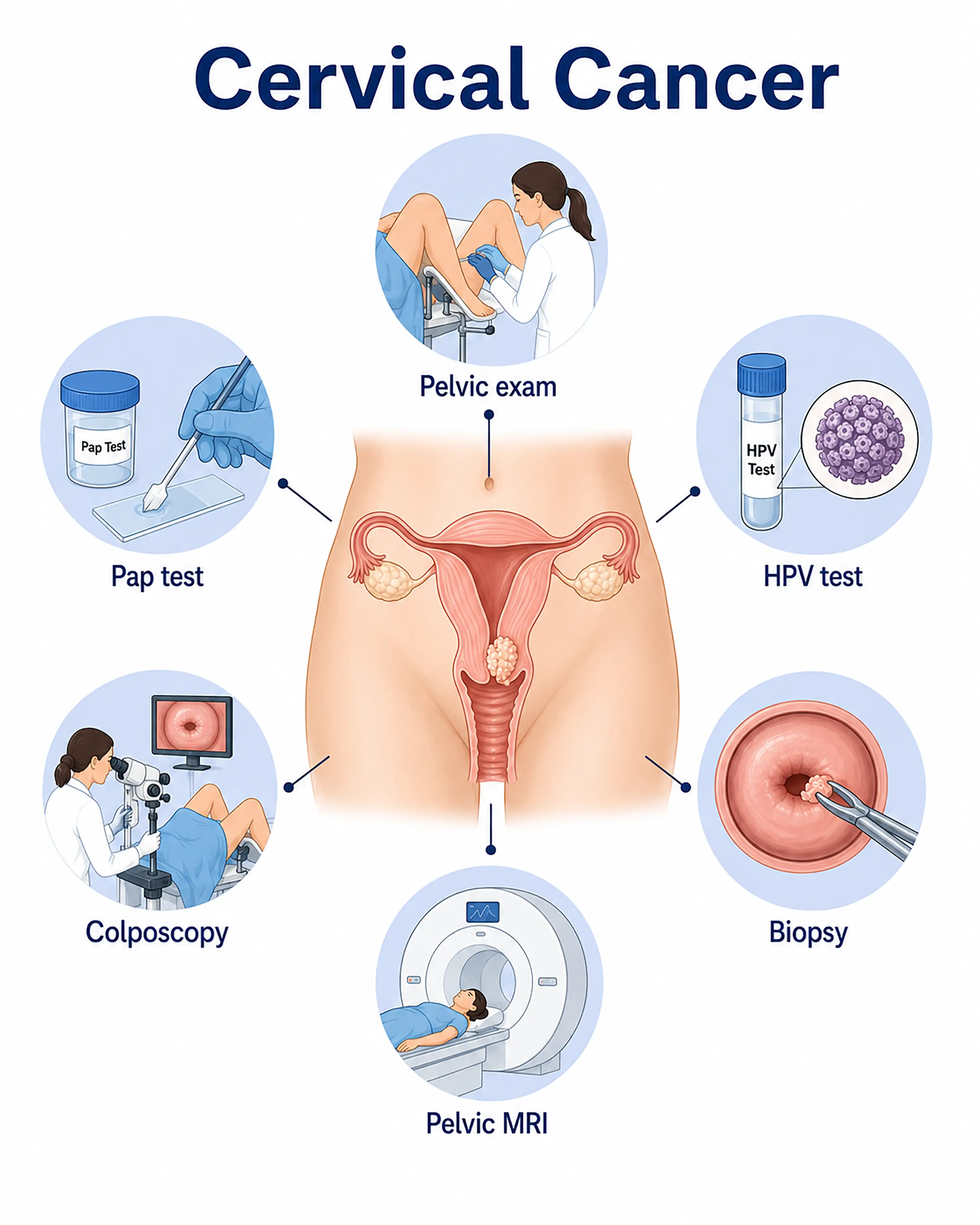
Cervical cancer diagnosis by stage
1.Early-stage cervical cancer diagnosis
Cancer cells remain within the cervical epithelium at a precancerous or carcinoma in situ stage. Combined TCT and HPV screening detects abnormalities; colposcopic acetic acid and iodine tests locate lesions, and biopsy confirms the diagnosis.
2.Intermediate-stage cervical cancer diagnosis
The tumor invades the cervical stroma, producing cauliflower-like or nodular enlargement. Cervical biopsy and endocervical curettage establish pathology, while pelvic MRI assesses tumor size and parametrial invasion.
3.Advanced-stage cervical cancer diagnosis
The tumor invades the vagina, parametrium, or pelvic wall, with regional nodal metastasis. Whole-body CT or PET-CT evaluates distant metastases, while biopsy establishes pathology to guide chemoradiotherapy.
Without timely diagnosis, cervical cancer continues to invade surrounding tissues and metastasize to lymph nodes and distant sites, potentially eliminating surgical options and markedly worsening prognosis.
When to Seek Medical Attention
Early warning signs include vaginal bleeding after intercourse, irregular intermenstrual bleeding, postmenopausal bleeding, and abnormal discharge. Please consult a doctor through "Chat with a Doctor" to support early detection. During the online consultation, the doctor will ask about symptoms and menstruation and guide you through cervical screening.
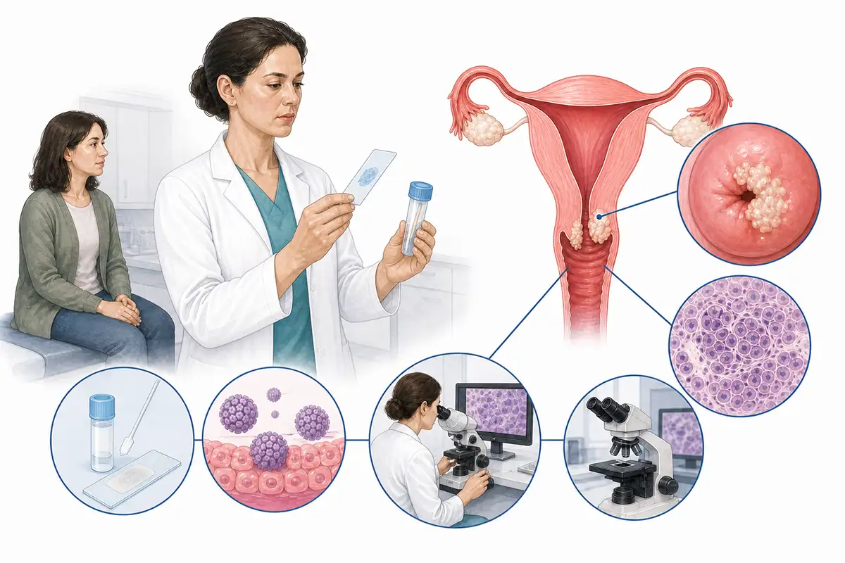
Basis for cervical cancer diagnosis
1.Cervical cytology
Thin-layer liquid-based cytology (TCT) collects cells from the cervical transformation zone to examine morphological changes. It is a basic cervical cancer screening method.
2.HPV testing
Testing detects high-risk HPV DNA, especially HPV16 and HPV18. Combined with TCT, it significantly improves detection of early lesions.
3.Colposcopy
Colposcopy magnifies vascular and morphological changes in cervical epithelium. Acetic acid and iodine tests locate suspicious areas and guide precise biopsy.
4.Cervical biopsy and pathology
Colposcopy-guided sampling of suspicious tissue for pathology is the gold standard for diagnosing cervical cancer and precancerous lesions and determines lesion grade.
5.Endocervical curettage
A specialized curette collects tissue from the cervical canal for pathology to assess endocervical disease, particularly when investigating adenocarcinoma.
6.Pelvic MRI
MRI clearly shows cervical tumor size, invasion depth, parametrial invasion, and vaginal involvement, outperforming CT for local staging.
7.Chest and abdominal CT
These examinations assess metastases in mediastinal lymph nodes, lungs, liver, and retroperitoneal nodes to establish distant spread.
8.Whole-body PET-CT assessment
Whole-body radioactive tracer imaging detects small metastases that conventional imaging may miss, allowing precise staging.
9.Cervical cancer screening recommendations
Sexually active women over 21 are advised to undergo regular combined TCT and HPV screening. HPV vaccination effectively prevents high-risk HPV infection.
Daily Self-Check
Monitor abnormal vaginal bleeding and discharge, and seek immediate care for postcoital or postmenopausal bleeding. Women in the appropriate age groups should receive regular combined TCT and HPV screening. HPV vaccination is an effective preventive measure.
Conclusion
Cervical cancer is currently the only malignancy with an established cause that can be detected early through screening. Standardized screening and early diagnosis significantly reduce mortality, while HPV vaccination is an important preventive measure.


