


Global disease prevalence: Cerebral arteriovenous malformation is an uncommon cerebrovascular disease worldwide; it can occur at any age and carries a high risk of danger following rupture and hemorrhage.
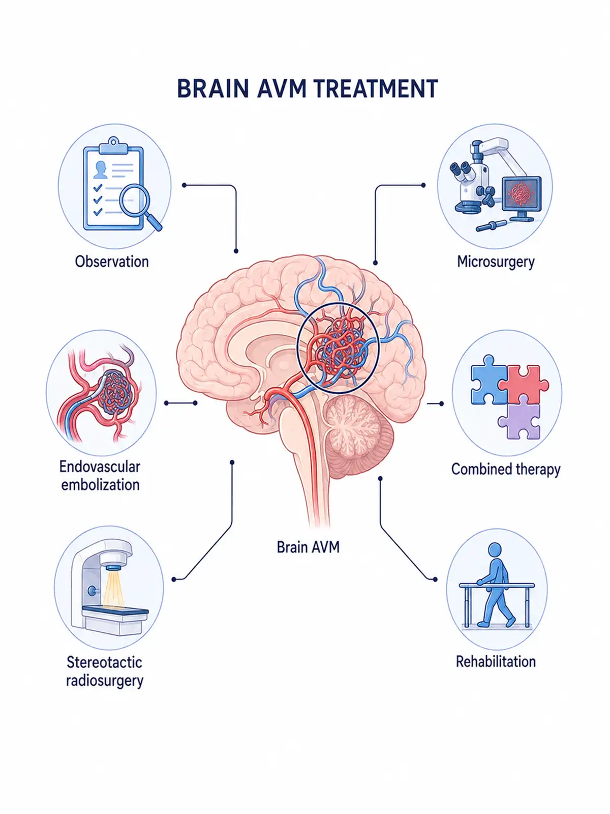
Staged treatment for cerebral arteriovenous malformation
Early treatment (involving localized lesions and reduced tumor volume for good results)
via surgical excision combined with adjuvant therapy can effectively control the risk of bleeding, leading to a relatively favorable prognosis.
Intermediate-stage treatment (addressing lesion enlargement, inhibiting progression, and delaying deterioration)
employs a comprehensive approach—combining surgery with embolization—to control further lesion enlargement, reduce the risk of hemorrhage, and slow disease progression.
Advanced-stage treatment (addressing severe hemorrhage, alleviating symptoms, and prolonging survival)
primarily employs systemic therapies such as embolization and radiotherapy to relieve neurological symptoms and minimize the risk of re-bleeding.
Consequences of delayed early treatment: Failure to receive timely, standardized treatment in the early stages may lead to the rupture of malformed blood vessels, causing severe cerebral hemorrhage; missing the optimal window for intervention can be life-threatening.
Why choose Fosun Health for cerebral arteriovenous malformation treatment
Fosun Health prioritizes the diagnosis and treatment of brain arteriovenous malformations (BAVM) within its neurosurgery and precision oncology services.The neurosurgery department at Foshan Fosun Chancheng Hospital—part of the Fosun Health network—was established in 1994; after nearly three decades of development, it is equipped with top-tier diagnostic technology, including dual-source CT, 3.0T MRI, DSA, and PET-CT systems.Department leader Professor Chen Baozhong has extensive experience in the surgical treatment of ruptured intracranial aneurysms and arteriovenous malformations, while experts such as Tan Feng and Wang Enmin specialize in CyberKnife radiosurgery for BAVM.
Leveraging Fosun Health’s global medical network and innovative R&D capabilities, patients have access to diverse treatment options.For cerebral arteriovenous malformations (AVMs) that are deep-seated, located in critical functional areas, or carry high surgical risk, the CyberKnife system at Foshan Fosun Chancheng Hospital enables precise stereotactic radiotherapy; for cases suitable for surgery, the neurosurgery team performs comprehensive treatments such as microsurgical resection and endovascular embolization.Additionally, FCN-159—a MAPK pathway inhibitor independently developed by Fosun Pharma—has entered the clinical research stage for treating cerebral AVMs, while Luvometinib (Fumaining®) has demonstrated an objective response rate of 64.3% (based on DSA assessment) in treating extracranial AVMs, offering new possibilities for targeted drug therapy in complex cases.
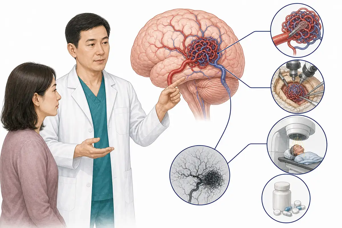
Treatment Methods for Cerebral Arteriovenous Malformation
1.Microsurgical resection of cerebral arteriovenous malformation
For cerebral arteriovenous malformations (AVMs) where location, size, and vascular anatomy are suitable for surgery, microsurgical resection of the malformed vascular nidus can be performed to directly eliminate the abnormal arteriovenous shunt.Some complex cases may be treated using a combination of modalities, such as preoperative endovascular embolization.
Treatment advantages:
① Direct management: Direct resection of the vascular malformation nidus can rapidly eliminate abnormal arteriovenous shunting.
② Immediate treatment: Directly treating the malformation nidus after surgery for lesions with clear surgical indications.
③ Applicable to cases with hemorrhage: For some patients who have experienced hemorrhage and meet surgical indications, comprehensive management addressing both the hematoma and the abnormal vascular nidus can be performed.
④ Personalized surgery: Formulate a surgical plan based on the size and location of the malformation and the vascular architecture (including feeding arteries and draining veins).
2.CyberKnife precision radiotherapy
For certain cerebral arteriovenous malformations (AVMs) that are deep-seated, located in critical functional areas, or carry a high risk for direct surgery, CyberKnife stereotactic radiotherapy may be considered based on lesion size and indications for radiotherapy.Obliteration of the malformed vessels typically requires time after treatment, and long-term follow-up via imaging is necessary.
Treatment advantages:
① Precise localization: Determine the location of the vascular malformation nidus using imaging, enhancing the accuracy of target area localization.
② Non-craniotomy treatment: Does not require traditional craniotomy; suitable for certain lesions not amenable to direct surgery.
③ Protects surrounding tissues: Uses stereotactic technology to concentrate radiation on the target area, minimizing the radiation dose to surrounding normal brain tissue.
④ Applicable to special sites: Can serve as a local treatment option for lesions located deep within the body or in areas of critical function.
3. VitalBeam linear accelerator precision radiotherapy
Utilizing the VitalBeam linear accelerator platform, precise irradiation—such as stereotactic radiotherapy—can be administered to selected eligible cerebral arteriovenous malformations.Specific treatment plans require a comprehensive assessment based on the size and location of the malformation and surrounding critical neural structures.
Treatment advantages:
① Precision irradiation: Formulate an individualized radiotherapy plan based on the location of the abnormal vascular nidus.
② Dose optimization: Concentrates the therapeutic dose in the target area by optimizing the radiation dose distribution.
③ Reduce radiation exposure to normal tissue: Minimize radiation exposure to surrounding normal brain tissue through precise planning.
④ Non-craniotomy treatment: It avoids the need for traditional craniotomy and serves as a local treatment option for some patients.
4.Endovascular interventional embolization
Employs a catheter to access the feeding arteries of a cerebral arteriovenous malformation (AVM) via the vasculature, using embolic materials to reduce or block abnormal blood flow.Depending on the angioarchitecture of the lesion, embolization may serve as a standalone treatment for some patients or be combined with microsurgery or radiotherapy.
Treatment advantages:
① Minimally invasive intervention: Treatment performed via a vascular route, eliminating the need for traditional craniotomy.
② Reduce abnormal shunting: Decrease blood flow to the malformation nidus by treating specific feeding vessels.
③ Combination therapy: Can be combined with surgery or radiotherapy based on the characteristics of the lesion.
④ Vascular architecture-guided: Interventional plans are formulated based on the feeding arteries, draining veins, and the structure of the malformation nidus.
5.Observation and follow-up
For selected small, asymptomatic cerebral arteriovenous malformations (AVMs) deemed unsuitable for immediate intervention upon professional assessment, a strategy of periodic observation may be adopted, monitoring changes in the lesion via imaging modalities such as MRI, MRA, or cerebral angiography.
6.Comprehensive therapy
A staged treatment plan—combining endovascular embolization, microsurgical resection, and stereotactic radiotherapy—may be formulated based on the size, location, angioarchitecture, and treatment risks of the vascular malformation nidus.
7.Antiepileptic therapy
For patients experiencing seizures, antiepileptic drugs may be used to control symptoms based on the seizure type and clinical condition, alongside comprehensive management that includes treatment of the underlying cerebrovascular malformation.
8.Rehabilitation therapy
For patients experiencing motor, speech, swallowing, or cognitive impairments following hemorrhage, surgery, or other treatments, targeted rehabilitation training may be implemented based on the specific functional deficits.
9.Supportive care
Address headaches, cerebral edema, and other related symptoms through appropriate symptom management, nutritional support, and psychological support tailored to the clinical situation.
Post-treatment care
Manage rest and activity levels according to medical advice; avoid strenuous exercise and behaviors that may increase the risk of bleeding; maintain proper blood pressure control; and adhere to the prescribed regimen for anti-epileptic and other medications.
Post-treatment examinations
Depending on the treatment method and the nature of the lesion, regular imaging examinations—such as MRI, MRA, or cerebral angiography—are required; these are combined with neurological function assessments to evaluate the occlusion status of the abnormal vascular nidus and the treatment efficacy.
Conclusion
Cerebral arteriovenous malformation poses a serious threat; successful treatment hinges on early detection, diagnosis, and intervention, as well as selecting an appropriate treatment plan and adhering to follow-up care to reduce the risk of hemorrhage.


