


Cerebral arteriovenous malformation (AVM) is a rare cerebrovascular lesion with an incidence of approximately 1–2 cases per 100,000 people.Most patients are born with the condition, though symptoms typically manifest between the ages of 10 and 40. Approximately 50% of AVM cases present initially with cerebral hemorrhage, and the annual risk of hemorrhage is about 2%–4%.
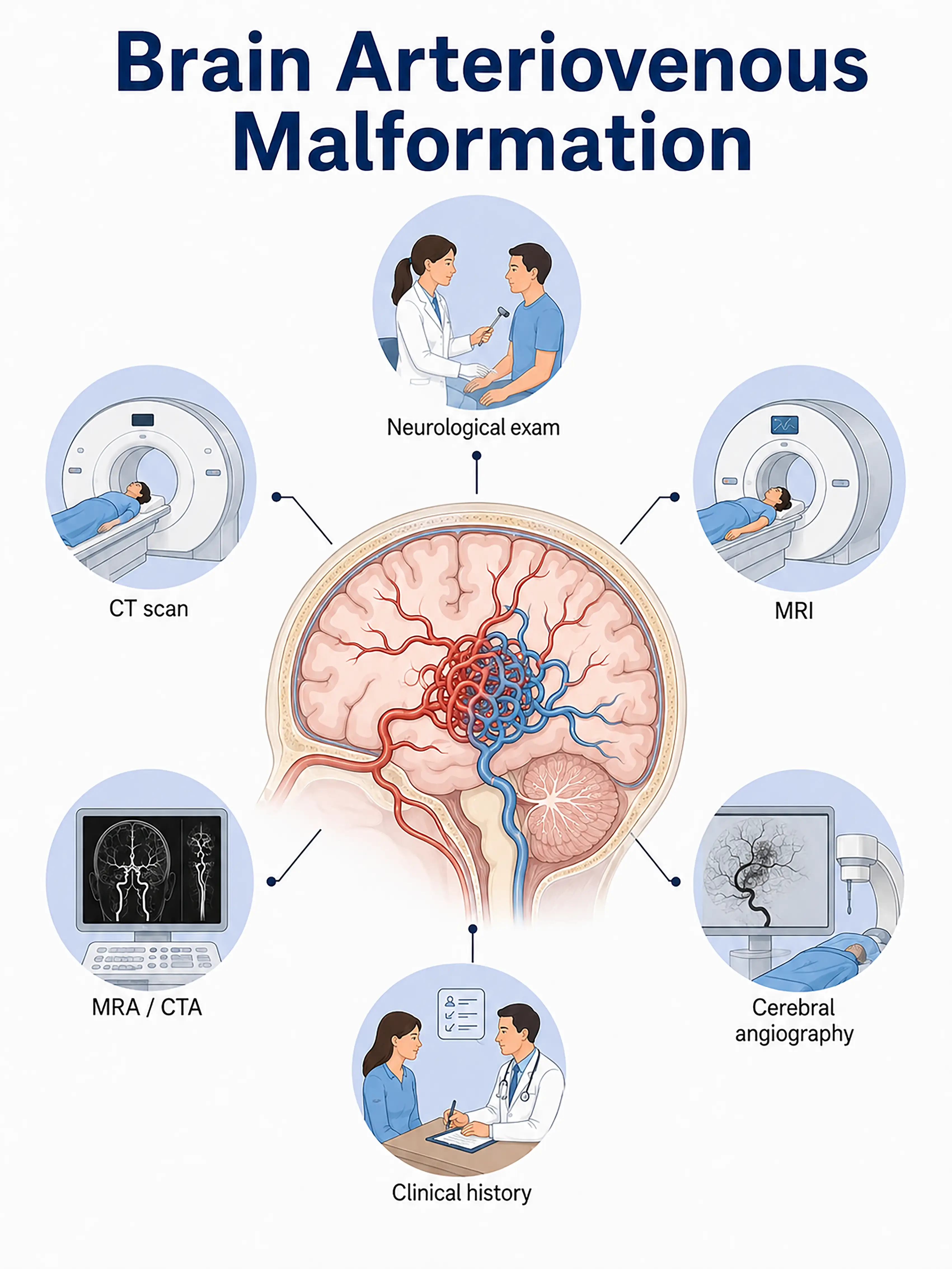
Diagnosis based on stages of cerebral arteriovenous malformation
1.Early diagnosis of cerebral arteriovenous malformation
Approximately 15% of AVMs remain asymptomatic for long periods and are discovered incidentally during physical examinations.MRI reveals a cluster of abnormal blood vessels with flow-void signals, and MRA can visualize feeding arteries and draining veins; definitive diagnosis requires DSA.
2.Diagnosis of mid-stage cerebral arteriovenous malformation
Seizures, headache, or progressive neurological deficits.MRI shows a characteristic "honeycomb" flow-void signal; DSA defines the size of the malformation nidus, feeding arteries, and draining veins.
3.Diagnosis of advanced cerebral arteriovenous malformation
Rupture of the malformed vessels leads to cerebral hemorrhage, presenting with severe headache, impaired consciousness, and hemiplegia.Emergency CT scans reveal the hemorrhage, while CTA or DSA confirms the diagnosis of AVM.
If a cerebral AVM is not diagnosed in time, rupture of the malformed vessels can lead to fatal intracerebral hemorrhage; the mortality rate for each hemorrhagic event is approximately 10%, and the cumulative risk increases with age.
When to Seek Medical Attention
Early signs of a cerebral arteriovenous malformation (AVM) include severe headaches, seizures, limb weakness, and speech difficulties.Please consult a doctor via the "Chat with a Doctor" feature for early detection.During the online consultation, the doctor will ask about your symptoms and family history and guide you through the process of obtaining cerebrovascular imaging.
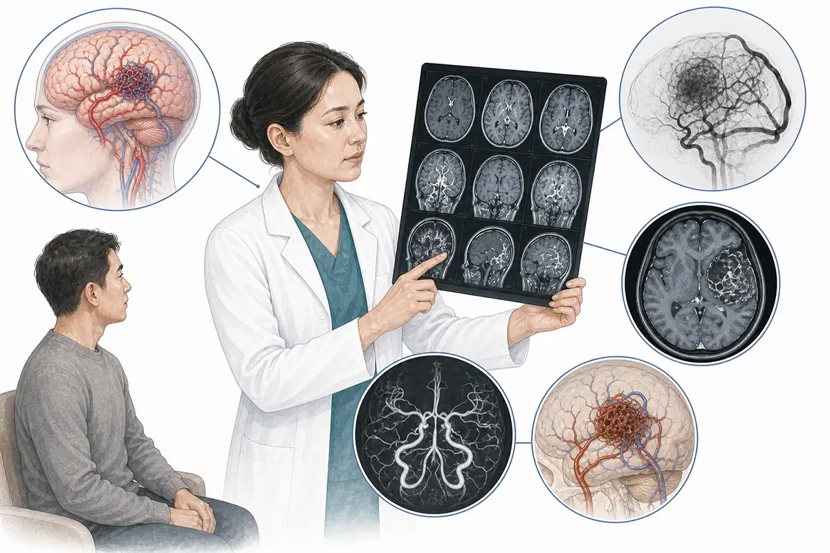
Diagnostic criteria for cerebral arteriovenous malformation
1.Digital subtraction angiography
DSA is the gold standard for diagnosing AVMs; it clearly displays the size and location of the nidus, as well as feeding arteries and draining veins, making it an essential examination for formulating treatment plans.
2.Cranial MRI (non-contrast and contrast-enhanced)
MRI typically reveals a "honeycomb" or "earthworm-like" flow-void signal pattern within the brain parenchyma.Contrast-enhanced MRI can visualize enhancing vascular structures and assess the relationship between the AVM and surrounding brain tissue.
3.CT angiography
CTA allows for rapid assessment of the vascular structure of an AVM—visualizing feeding arteries, the nidus, and draining veins—and is the preferred examination in emergency settings.
4.MRA examination
Non-invasive assessment of AVM vascular structure—while offering lower spatial resolution than DSA—is suitable for preoperative evaluation and follow-up monitoring, thereby reducing the frequency of DSA examinations.
5.Spetzler-Martin grading
A grading system (Grades 1–5) based on AVM size (<3 cm, 3–6 cm, >6 cm), location (eloquent/non-eloquent cortex), and venous drainage (deep/superficial) is used to guide treatment decisions.
6.Functional brain MRI
fMRI evaluates the spatial relationship between AVMs and critical functional areas (motor and language zones), guiding surgical planning and reducing the risk of postoperative neurological deficits.
7.Diffusion tensor imaging
DTI visualizes the relationship between the AVM and white matter tracts and assesses the potential impact of surgery on neural conduction pathways, serving as a valuable supplement to preoperative evaluation.
8.Transcranial Doppler ultrasound
Transcranial Doppler (TCD) detects changes in cerebral blood flow velocity and direction to assess the hemodynamic characteristics of AVMs, aiding in diagnosis and follow-up.
9.Brain perfusion imaging
CT or MR perfusion imaging assesses blood flow perfusion in the brain tissue surrounding an AVM, identifies the "steal phenomenon," and guides treatment strategies.
Daily Self-Check
Seek immediate medical attention if unexplained seizures, persistent headache, or cerebral hemorrhage occurs.Individuals with a family history of AVM (arteriovenous malformation) should undergo regular cerebrovascular examinations.
Conclusion
Diagnosis of cerebral AVMs relies on DSA (the gold standard) and MRI.The Spetzler-Martin grading scale guides treatment decisions, and a multidisciplinary approach represents the optimal model for diagnosis and care.


