


Cerebral AVMs are lesions resulting from abnormal cerebral vascular development during the embryonic stage.While arteries normally connect to veins via a capillary network, in an AVM, arteries and veins communicate directly, forming an abnormal vascular nidus characterized by high flow and low resistance.
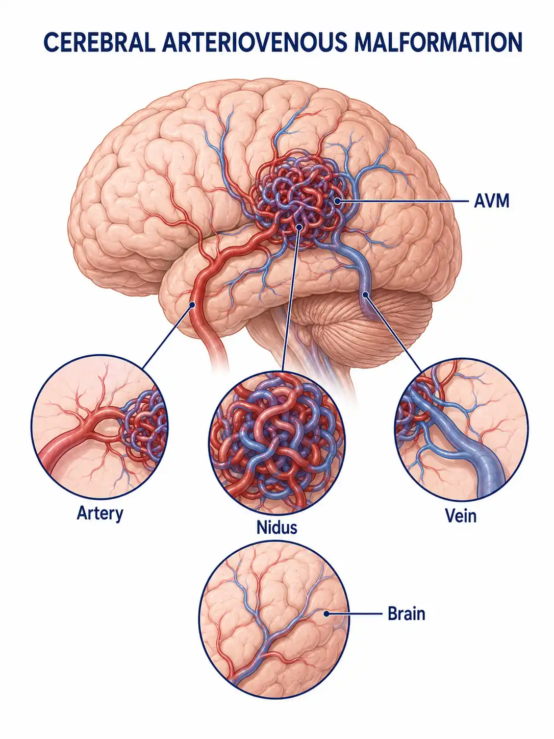
Global overview of cerebral arteriovenous malformation
The prevalence of cerebral arteriovenous malformation in the population is approximately 0.01%–0.5%. It is a significant cause of cerebral hemorrhage in young people, and the risk of hemorrhage increases with age.
Consult a doctor regarding cancer-related issues
Individuals presenting with sudden severe headache, seizures, progressive neurological deficits, or a family history of arteriovenous malformation (AVM) should promptly consult a neurosurgeon or interventional neuroradiologist.
Main types of cerebral arteriovenous malformation
1. Parenchymal AVM
The malformed vascular nidus is located within the brain parenchyma; this is the most common type and is prone to causing cerebral hemorrhage and epilepsy.
2. Periventricular AVM
The malformed vascular nidus is located in the ventricular wall or periventricular region; upon hemorrhage, it tends to rupture into the ventricle, causing intraventricular hemorrhage.
3. Dural AVM
Both the feeding arteries and draining veins are dural vessels; the condition is frequently associated with venous sinus thrombosis.
4. Brainstem AVM
Located in the brainstem region, these are deep-seated lesions with extremely high treatment risks; they represent the most difficult type of AVM to manage.
5. Diffuse AVM
The vascular malformation is extensive, involving multiple cerebral lobes; complete surgical resection is difficult, making treatment challenging.
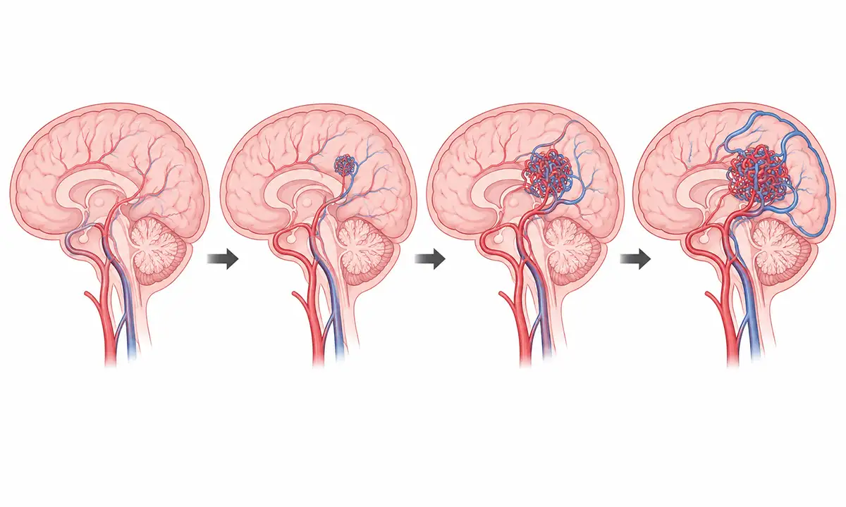
Typical symptoms and signs of cerebral arteriovenous malformation
1. Cerebral hemorrhage
Cerebral hemorrhage resulting from the rupture of the malformed vessels is the most severe complication, manifesting as sudden, severe headache, vomiting, and impaired consciousness.
2. Epileptic seizure
Irritation and compression of surrounding brain tissue by the malformed vascular nidus can trigger epileptic seizures, a common clinical manifestation of AVM.
3. Headache
Persistent or recurrent headache, which may be accompanied by a throbbing sensation, is associated with hemodynamic changes in abnormal blood vessels.
4. Neurological deficit
Progressive limb weakness, sensory disturbances, or speech difficulties; associated with ischemia of surrounding brain tissue caused by the "steal phenomenon."
5. Intracranial bruit
Patients with large arteriovenous malformations (AVMs) may hear a vascular bruit in the head that is synchronous with the pulse, indicating high-flow shunting.
6. Cognitive impairment
Large arteriovenous malformations (AVMs) in the frontal or temporal lobe can cause memory loss, inattention, and personality changes.
Diagnostic methods for cerebral arteriovenous malformation
1. Cranial CT
CT can detect cerebral hemorrhage and calcification, while contrast-enhanced scanning can provide a preliminary view of abnormal vascular clusters.
2. Cranial MRI
MRI clearly demonstrates the location and size of the vascular malformation nidus and its relationship with surrounding brain tissue; flow-void signals are a characteristic finding.
3. Cerebral angiography
DSA is the gold standard for diagnosing AVMs, capable of clearly defining the anatomical structures of feeding arteries, the nidus, and draining veins.
4. MRA examination
Magnetic resonance angiography (MRA) allows for the non-invasive visualization of the vascular structure of arteriovenous malformations (AVMs) and serves as an important tool for follow-up.
5. Functional assessment
fMRI and the Wada test assess the relationship between AVMs and critical functional areas, providing a basis for treatment decisions.
6. Grading assessment
The Spetzler-Martin grading system assesses surgical risk based on the size, location, and venous drainage pattern of an arteriovenous malformation (AVM).
Treatment Methods for Cerebral Arteriovenous Malformation
1. Microsurgery
Complete resection of an AVM via craniotomy is a curative treatment method, suitable for AVMs located superficially and in non-functional areas.
2. Interventional embolization
Endovascular embolization of the malformed vascular nidus can be used as a standalone treatment for small arteriovenous malformations (AVMs) or as an adjunct to surgery.
3. Stereotactic radiotherapy
Radiotherapy techniques such as Gamma Knife induce gradual occlusion of malformed vessels; this approach is suitable for small, deep-seated AVMs, though it takes 1–3 years to become effective.
4. Multimodal therapy
For large, complex arteriovenous malformations (AVMs), combined treatment regimens involving embolization plus surgery or embolization plus radiotherapy are employed.
5. Conservative treatment
For asymptomatic AVMs with a low risk of hemorrhage, a strategy of observation and follow-up with periodic MRI monitoring may be adopted.
6. Antiepileptic therapy
For patients with concurrent epilepsy, antiepileptic drugs are used to control seizures.
7. Rehabilitation therapy
Patients with residual neurological deficits following hemorrhage undergo rehabilitation training to improve motor and cognitive functions.
8. Follow-up monitoring
Periodic follow-up with MRA or DSA after treatment is essential to confirm the complete occlusion of the malformed vessels; long-term monitoring is crucial.
Conclusion
Cerebral arteriovenous malformation is a congenital cerebrovascular developmental anomaly, with cerebral hemorrhage being the most serious complication.The risk of hemorrhage can be effectively eliminated through methods such as microsurgery, interventional embolization, and stereotactic radiotherapy.


