


Cerebral arteriovenous malformation (AVM) is a lesion resulting from abnormal cerebrovascular development; arterial blood flows directly into veins without passing through capillaries, potentially leading to symptoms such as cerebral hemorrhage, seizures, and neurological deficits.
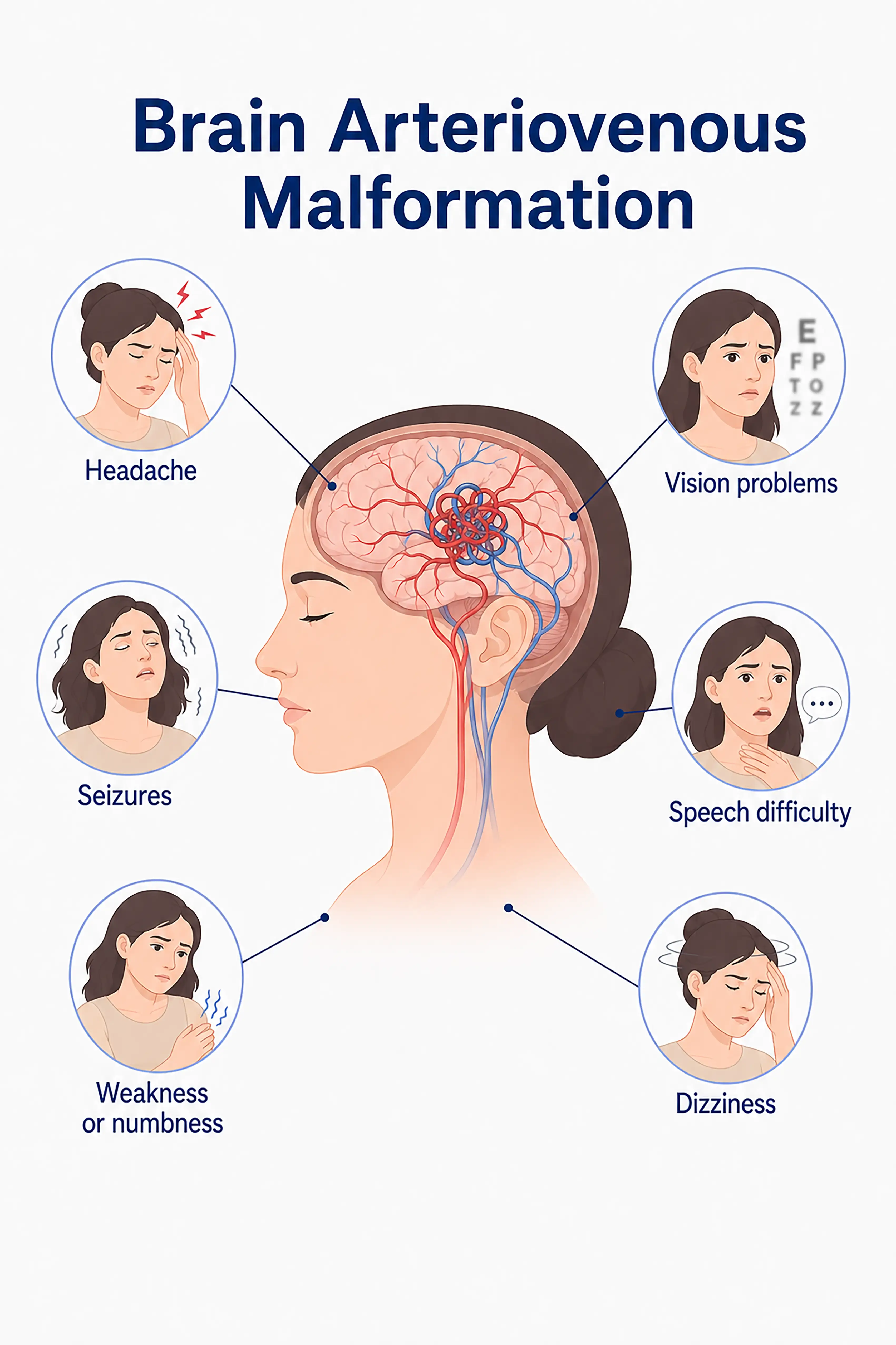
Prevalence of cerebral arteriovenous malformation worldwide
Cerebral arteriovenous malformation (AVM) is a rare cerebrovascular lesion with an incidence of approximately 1–2 cases per 100,000 people.While present at birth, symptoms usually manifest between the ages of 10 and 40. Approximately 50% of AVM cases present initially with cerebral hemorrhage, and the annual risk of hemorrhage is about 2%–4%.Patients with a history of prior hemorrhage face a higher risk of re-bleeding.
Symptoms associated with stages of cerebral arteriovenous malformation
1.Early symptoms of cerebral arteriovenous malformation
Approximately 15% of AVMs remain asymptomatic for long periods and are discovered incidentally during physical examinations.Symptomatic cases may present with mild headaches, transient dizziness, or mild cognitive difficulties, which are easily overlooked.
2.Symptoms of mid-stage cerebral arteriovenous malformation
Persistent headache, seizures, progressive muscle weakness, sensory abnormalities, speech impairment, and vision problems occur; symptoms gradually worsen, affecting daily life.
3.Symptoms of advanced cerebral arteriovenous malformation
Rupture of the malformed vessels leads to massive hemorrhage, resulting in severe headache, loss of consciousness, hemiplegia, coma, or even life-threatening consequences.Recurrent hemorrhages can cause severe neurological impairment.
When to Seek Medical Attention
Early signs of a cerebral arteriovenous malformation (AVM) include severe headaches, seizures, limb weakness, and speech difficulties.Please consult a doctor via the "Chat with a Doctor" feature for early detection.During the online consultation, the doctor will ask about your symptoms and family history and guide you through the process of obtaining cerebrovascular imaging.
Staging of cerebral arteriovenous malformation
Cerebral Arteriovenous Malformation: Stage 0
This staging system is not applicable to cerebral arteriovenous malformations (AVMs).AVMs are graded using the Spetzler-Martin grading system, based on size, location, and venous drainage.
Cerebral Arteriovenous Malformation: Stage I
Small AVM (diameter <3 cm) located in a non-eloquent area with deep venous drainage; associated with a relatively low risk of hemorrhage.
Cerebral Arteriovenous Malformation: Stage II
Medium-sized AVM (diameter 3–6 cm) located near critical functional areas; symptoms appear gradually, with risks of hemorrhage and epilepsy.
Cerebral Arteriovenous Malformation: Stage III
Large AVMs (diameter >6 cm) located in functional areas with deep venous drainage present severe symptoms and are difficult to treat.
Cerebral Arteriovenous Malformation: Stage IV
A giant, complex arteriovenous malformation (AVM) located in the brainstem or deep structures; it carries an extremely high risk of hemorrhage and can lead to severe neurological impairment and death.
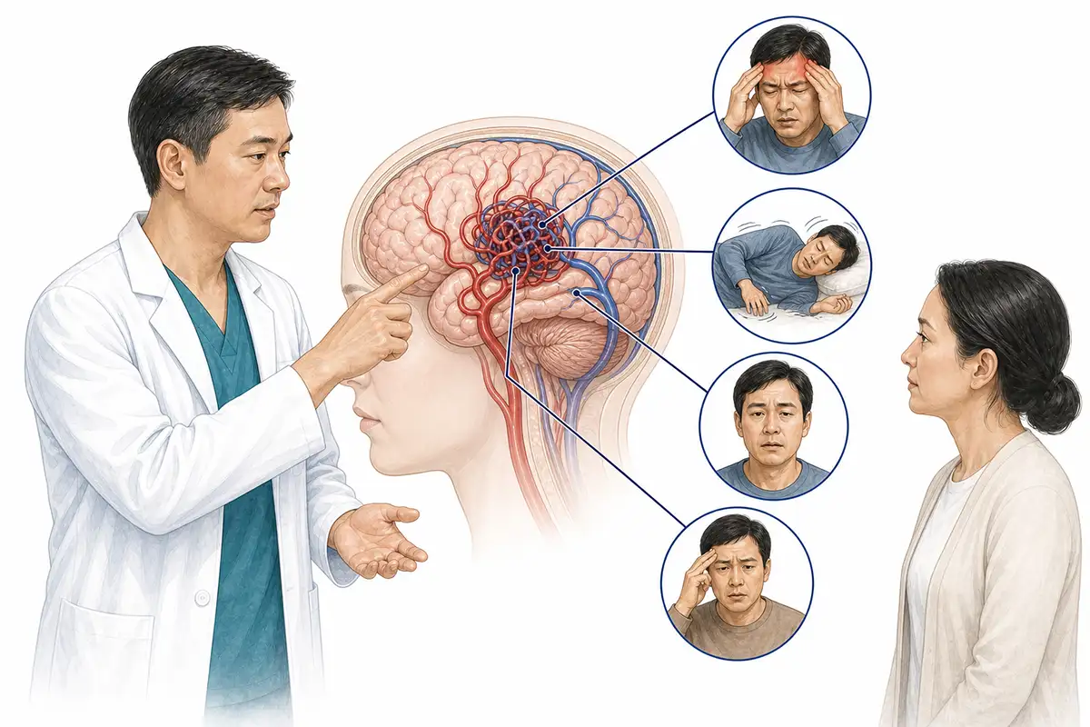
Detailed Symptoms of Cerebral Arteriovenous Malformation
1.Onset of cerebral hemorrhage
In more than 50% of AVM cases, the initial symptom is cerebral hemorrhage; rupture of the malformed vessels leads to sudden severe headache, nausea and vomiting, impaired consciousness, and neurological deficits.
2.Epileptic seizures
Approximately 20%–25% of patients present with seizures as the initial symptom; irritation of brain tissue and ischemia caused by the AVM can lead to focal or generalized seizures.
3.Persistent headache
High blood flow through an AVM can cause persistent headaches—resembling migraines or throbbing pain—ranging from mild to severely debilitating.
4.Limb weakness/numbness
AVM-associated "steal phenomenon" causes ischemia and hypoxia in surrounding brain tissue, resulting in reduced limb muscle strength, numbness, tingling, or sensory loss.
5.Speech impairment
When an AVM is located in a language-related area or hemorrhage involves the language center, difficulties in speech expression or comprehension deficits occur.
6.Visual problems present
Occipital lobe AVMs or hemorrhages involving the visual pathways result in visual field defects, blurred vision, or ocular motility disorders.
7.Decline in cognitive memory
An AVM affecting frontal or temporal lobe function leads to impaired attention, memory loss, and executive dysfunction.
8.Presence of unsteady gait
Cerebellar or brainstem AVM causing balance disorders, ataxia, and walking difficulties, accompanied by dizziness and impaired coordination.
9.Intracranial bruit present
Some patients with large arteriovenous malformations (AVMs) may hear a pulsatile murmur in the head that is synchronized with the heartbeat; this is caused by high-velocity blood flow.
Conclusion
The goals of treating a cerebral AVM are to eliminate the risk of hemorrhage and prevent seizures.Treatment options include microsurgery, stereotactic radiotherapy, and endovascular embolization; the choice of treatment requires a comprehensive assessment based on the Spetzler-Martin grading scale and the patient's condition.


