


Anal cancer is a rare malignancy of the digestive tract, accounting for approximately 2% of cases.It is more common in women than in men.About 90% of cases are associated with HPV infection (particularly HPV-16).Squamous cell carcinoma of the anal canal is the most common histological type.The incidence is higher among individuals with HIV infection and those who are immunosuppressed.
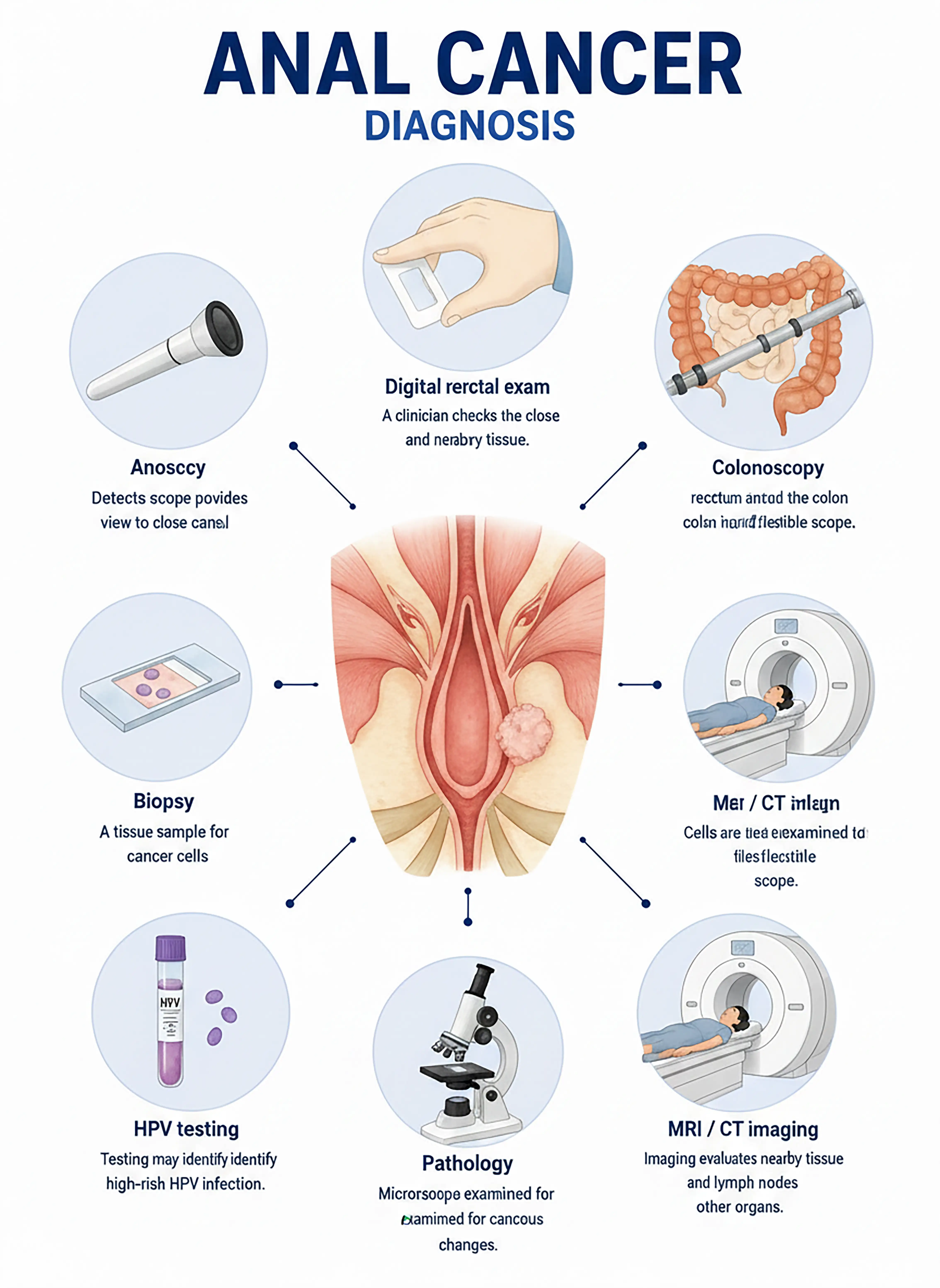
Staging and diagnosis of anal cancer
1.Early diagnosis of anal cancer
The tumor is confined to the anal canal mucosa, presenting with anal bleeding or itching.Anoscopy reveals mucosal abnormalities, and biopsy establishes the pathological diagnosis.
2.Diagnosis of mid-stage anal cancer
Presentation includes anal pain and a palpable mass.Anoscopy-guided biopsy is used to determine the pathological type, while contrast-enhanced MRI assesses tumor extent and inguinal lymph node metastasis.
3.Diagnosis of advanced anal cancer
Tumor invasion of the rectum or vagina, or distant metastasis to the liver and lungs.Whole-body CT and PET-CT are used to assess the extent of metastasis.
If not diagnosed promptly, the tumor may invade adjacent organs and metastasize distantly, potentially requiring abdominoperineal resection (APR), which severely impacts quality of life.
When to Seek Medical Attention
Early signs of anal cancer include anal bleeding, pain, itching, and lumps.Please consult a doctor via "Chat with a Doctor" for early detection.During the online consultation, the doctor will ask about your symptoms and history of HPV infection, and guide you through an anoscopy.
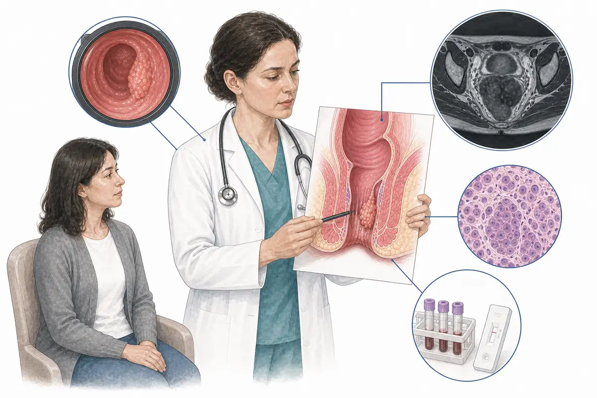
Basis for diagnosing anal cancer
1.Anoscopy
An anoscope is inserted into the anal canal to directly observe the tumor's location, size, and morphology, and to perform a biopsy under visualization; it is a key tool for diagnosing anal cancer.
2.Histopathological biopsy
Obtaining a biopsy of suspicious tissue under anoscopy guidance for pathological examination is the gold standard for diagnosing anal cancer, allowing for the determination of the pathological type and degree of differentiation.
3Pelvic MRI
Contrast-enhanced MRI is the preferred imaging modality for assessing the extent of local invasion in anal cancer, clearly demonstrating the relationship between the tumor and the anal sphincter and surrounding tissues.
4.Digital rectal examination
A physician inserts a gloved finger into the anus to palpate the anal canal and rectum for masses, ulcers, or strictures; this is a simple yet important preliminary examination.
5.HPV testing
Detecting HPV infection status (particularly HPV type 16)—as approximately 90% of anal cancers are associated with HPV infection—aids in etiological analysis and prognostic assessment.
6.Inguinal lymph node assessment
Palpation or ultrasound is used to assess for inguinal lymph node enlargement, guide fine-needle aspiration biopsy of suspicious nodes, and evaluate metastatic status.
7.Chest and abdominal CT
Assesses for distant metastases in the lungs, liver, and retroperitoneal lymph nodes; a standard examination for initial staging.
8.HIV testing
Patients with anal cancer should undergo routine HIV testing; the incidence of anal cancer is high among HIV-infected individuals, and immune status influences the choice of treatment regimen.
9.Whole-body PET-CT
Assess for distant metastasis; offers advantages in detecting occult metastatic lesions that are difficult to identify via conventional imaging, aiding in precise staging.
Daily Self-Check
Seek medical attention promptly if symptoms such as anal bleeding, pain, itching, or a palpable mass occur.Individuals with HPV or HIV infections should undergo regular anal screening.
Conclusion
Diagnosis of anal cancer relies on anoscopy and biopsy. HPV infection is a major risk factor;early detection allows for anal-sparing treatment via chemoradiotherapy.


