


Adrenocortical carcinoma is a very rare malignant tumor with an annual incidence of approximately one in a million.It most commonly affects individuals aged 40–50, with a slightly higher incidence in women.About 60% are functional tumors capable of secreting cortisol, androgens, or aldosterone.Tumors often exceed 6 cm in diameter, and the prognosis is generally poor.
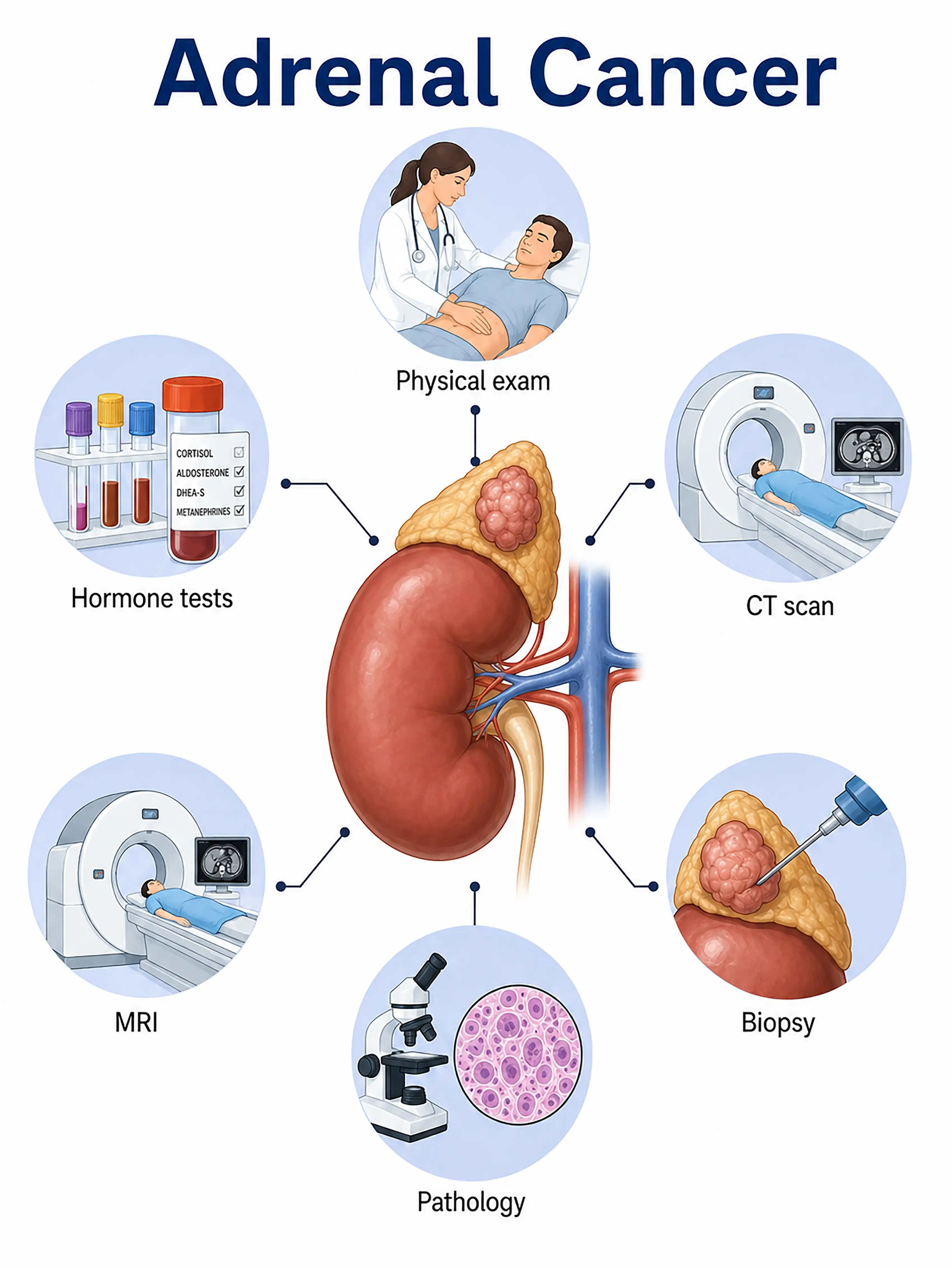
Diagnosis of adrenocortical carcinoma by stage
1.Early diagnosis of adrenal cancer
An adrenal mass (>4 cm) is incidentally discovered via imaging.CT shows heterogeneous enhancement and a delayed washout rate of <58%; hormonal testing assesses functional status, and diagnosis is confirmed via needle aspiration or surgical biopsy.
2.Diagnosis of mid-stage adrenal cancer
Symptoms of hormone excess (Cushing's syndrome, virilization) or an abdominal mass.Contrast-enhanced CT and MRI are used to assess tumor extent and vascular invasion;biopsy confirms the pathology.
3.Diagnosis of advanced adrenal cancer
Tumor extensively invades surrounding organs or metastasizes to the lungs and liver.Whole-body CT and PET-CT are used to assess the extent of metastasis, and surgical acquisition of a complete specimen is required to determine the stage.
If not diagnosed promptly, functional tumors cause severe endocrine disorders, while non-functional tumors are often at an advanced stage upon detection, resulting in a very poor prognosis.
When to Seek Medical Attention
Early signs of adrenocortical carcinoma include an abdominal mass, unexplained weight gain, abnormally high blood pressure, and purple skin striae. Please consult a doctor via the "Chat with a Doctor" feature to facilitate early detection.During the online consultation, the doctor will ask about your symptoms and hormonal changes, and guide you through imaging studies and hormone tests.
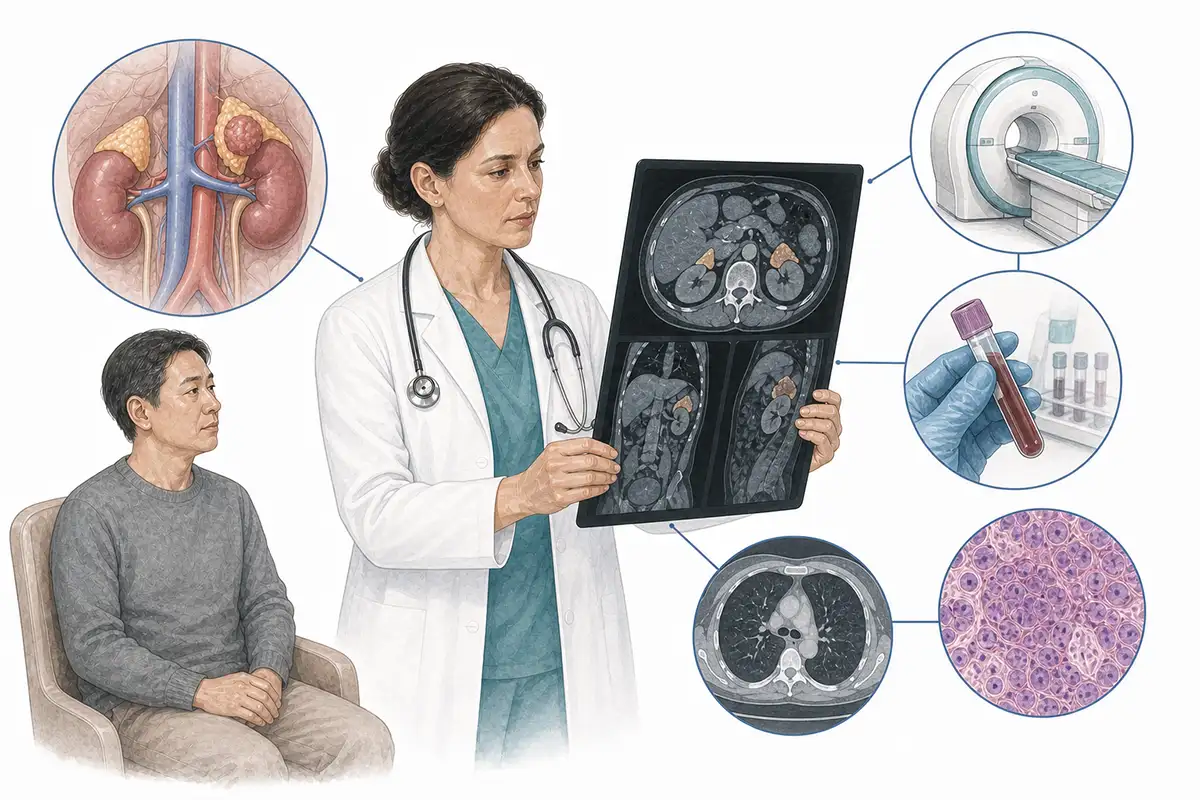
Basis for Diagnosing Adrenocortical Carcinoma
1.Contrast-enhanced CT scan
Contrast-enhanced CT is the preferred imaging modality for diagnosing adrenocortical carcinoma; typical features include a tumor size >4 cm, heterogeneous enhancement, ill-defined margins, and a delayed washout rate of <58%.
2.Abdominal MRI scan
MRI shows isointense or hypointense signals on T1-weighted images and hyperintense signals on T2-weighted images, with signal loss observed on chemical shift imaging; it allows for the assessment of the tumor's relationship with surrounding blood vessels and organs.
3.Comprehensive hormonal assessment
Test for 24-hour urinary free cortisol, plasma ACTH, androgens, aldosterone, and catecholamines to determine the functional status of the tumor and rule out pheochromocytoma.
4.Histopathological examination
Pathological examination of the surgically resected specimen, assessing malignancy via the Weiss scoring system, serves as the gold standard for definitive diagnosis.
5.Dexamethasone suppression test
The high-dose dexamethasone suppression test assesses whether cortisol secretion is suppressed, aiding in the determination of the tumor's functional status and in differential diagnosis.
6.Chest CT scan
Assesses for pulmonary metastases; approximately 40%–80% of metastatic cases of adrenocortical carcinoma involve the lungs.
7.PET-CT assessment
18F-FDG PET-CT demonstrates high metabolic activity, assesses distant metastasis and local recurrence, and assists with staging and treatment efficacy evaluation.
8.Tumor size assessment
The likelihood of malignancy increases significantly when the tumor diameter exceeds 4 cm; a diameter greater than 6 cm indicates malignancy with 91% sensitivity and 80% specificity.
9.Genetic testing analysis
Detects mutations in genes such as *TP53* (associated with Li-Fraumeni syndrome) and *PRKAR1A*, assisting in screening for genetic syndromes and molecular subtyping.
Daily Self-Check
Seek medical attention promptly if an unexplained abdominal mass, signs of Cushing's syndrome, or abnormal blood pressure occurs.Individuals with an adrenal mass should undergo regular hormonal assessment.
Conclusion
Diagnosis relies on CT/MRI and hormone testing.Early detection and radical surgical resection are key to improving prognosis.


