


Multiple myeloma is a hematologic malignancy characterized by the malignant clonal proliferation of plasma cells.Malignant plasma cells proliferate extensively in the bone marrow and produce monoclonal immunoglobulins, leading to osteolytic bone destruction, anemia, renal impairment, and immune dysfunction.It predominantly affects middle-aged and elderly individuals, with a slightly higher incidence in men than in women, and is the second most common hematologic malignancy.
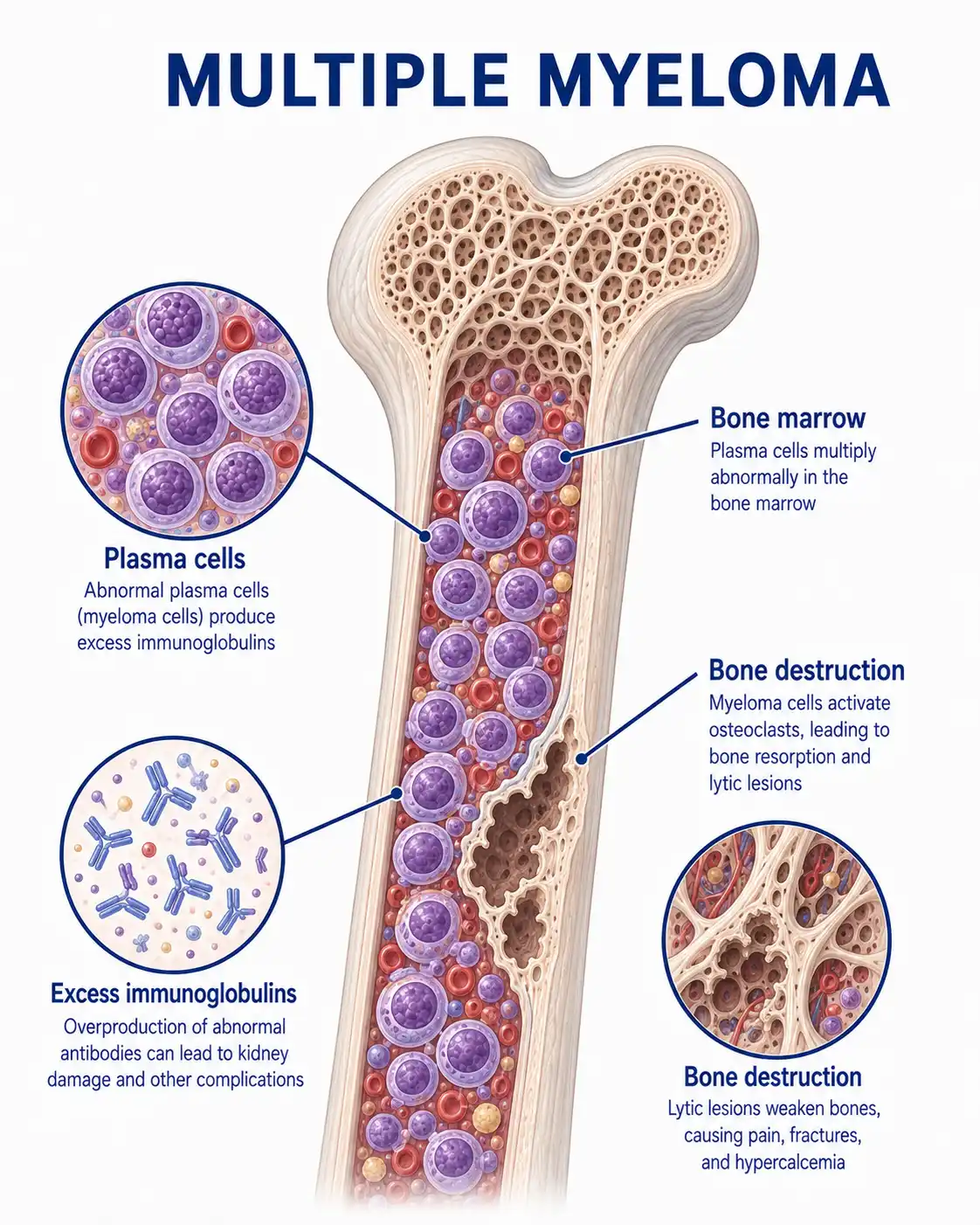
Global overview of multiple myeloma
The global annual incidence of multiple myeloma is approximately 4–5 cases per 100,000 people, accounting for over 10% of hematologic malignancies. Incidence rates are higher in developed countries than in developing countries;the rate is highest among people of African descent and relatively lower among Asians. The peak age of onset is between 60 and 70 years;in recent years, the incidence has shown an upward trend due to population aging and improved diagnostic capabilities.
Consult a doctor regarding cancer-related issues
Middle-aged and elderly patients presenting with persistent bone pain (particularly in the lower back), recurrent infections, unexplained anemia, or abnormal renal function should promptly consult a hematologist.Upon diagnosis, they should undergo comprehensive assessment and receive personalized treatment at a specialized hematology department.
Main types of multiple myeloma
1.IgG type
The most common type, accounting for approximately 50%–60% of cases; it involves monoclonal IgG immunoglobulin and presents with classic clinical manifestations, including bone disease, anemia, and renal impairment.
2.IgA type
The second most common type, accounting for approximately 20%–25% of cases; the monoclonal immunoglobulin is IgA. Patients are more prone to hyperviscosity syndrome and extramedullary lesions, and the prognosis is relatively poor.
3.Light chain type
Accounts for approximately 15%–20% of cases; produces only monoclonal light chains and tests positive for urinary Bence-Jones protein. It carries a higher risk of renal impairment and amyloidosis, requiring aggressive hydration and chemotherapy.
4.IgD type
A rare subtype accounting for approximately 2% of cases; it is highly malignant and rapidly progressive, prone to extramedullary infiltration and amyloidosis, carries a poor prognosis, and requires aggressive treatment.
5.Non-secretory type
Accounts for approximately 1%–5% of cases; characterized by malignant proliferation of plasma cells in the bone marrow without the production of detectable monoclonal immunoglobulin. Diagnosis relies on bone marrow examination and tissue biopsy.
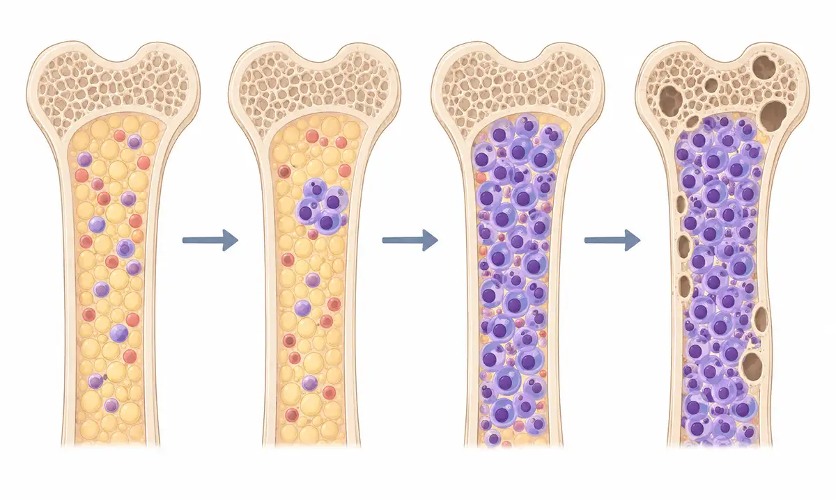
Typical symptoms and signs of multiple myeloma
1.Bone pain and fractures
Osteolytic lesions cause persistent bone pain—most commonly in the lower back, chest, and ribs—and can lead to pathological fractures from minor trauma, severely impairing mobility.
2.Anemia and fatigue
Suppression of normal bone marrow hematopoiesis leads to anemia, manifesting as pallor, fatigue, weakness, palpitations and shortness of breath upon exertion, and dizziness;symptoms progressively worsen as the disease advances.
3.Renal function impairment
Factors such as renal tubular damage caused by free light chain deposition, hypercalcemia, and dehydration lead to renal function impairment, manifesting as oliguria, edema, and elevated serum creatinine levels.
4.Recurrent infection
A reduction in normal immunoglobulins leads to impaired humoral immunity, increasing susceptibility to recurrent bacterial pneumonia and urinary tract infections; this is a significant cause of morbidity and mortality.
5.Hypercalcemia
Bone destruction releases large amounts of calcium ions into the bloodstream, resulting in symptoms of hypercalcemia such as nausea, vomiting, polyuria, constipation, confusion, and cardiac arrhythmias.
6.Symptoms of hyperviscosity
High concentrations of monoclonal immunoglobulin increase blood viscosity, leading to symptoms such as dizziness, blurred vision, tinnitus, a tendency to bleed, and retinal vein dilation.
Diagnostic methods for multiple myeloma
1.Complete blood count (CBC)
Shows normocytic, normochromic anemia with rouleaux formation of red blood cells; white blood cell and platelet counts may be reduced. Assess the severity of anemia and the extent of cytopenia.
2.Serum protein electrophoresis
Detection of serum M-protein bands to identify the type and concentration of monoclonal immunoglobulin; a crucial marker for diagnosis and monitoring treatment efficacy.
3.Immunofixation electrophoresis
Determine the heavy and light chain types of the M-protein—differentiating between IgG, IgA, IgM, IgD, and light-chain types—to assist in precise subtyping and diagnosis.
4.Free light chain assay
Measure the serum free light chain ratio; an abnormal ratio indicates the production of monoclonal light chains and serves as a sensitive indicator for early diagnosis and treatment efficacy assessment.
5.Bone marrow aspiration and biopsy
Bone marrow smears and biopsies showing an increased proportion of plasma cells with morphological abnormalities provide a key basis for diagnosis;flow cytometry can assist in detecting abnormal plasma cells.
6.Skeletal imaging examinations
Whole-body low-dose CT, MRI, or PET-CT assesses the extent and severity of osteolytic lesions, detecting early-stage bone destruction that conventional X-rays cannot identify.
Treatment Methods for Multiple Myeloma
1.Proteasome inhibitors: Proteasome inhibitors —such as bortezomib, carfilzomib, and ixazomib—induce apoptosis in tumor cells by inhibiting proteasome activity and serve as cornerstone drugs in the treatment of multiple myeloma.
2.Immunomodulatory agents: Immunomodulatory agents such as lenalidomide and pomalidomide possess multiple effects—including direct anti-tumor activity, immunomodulation, and inhibition of angiogenesis—and yield significant results when combined with proteasome inhibitors.
3.Monoclonal antibodies: Monoclonal antibodies such as the anti-CD38 agents daratumumab and elotuzumab can directly target and kill myeloma cells, significantly improving the depth of remission and survival.
4.Chemotherapy: Traditional chemotherapeutic agents, such as cyclophosphamide and melphalan, can be used for both newly diagnosed and relapsed patients; they are often combined with novel agents in multi-drug regimens.
5.Autologous hematopoietic stem cell transplantation:Autologous hematopoietic stem cell transplantation is suitable for patients whose age and physical condition permit;collecting autologous stem cells after induction chemotherapy, followed by high-dose chemotherapy combined with autologous transplantation, can prolong progression-free survival.
6.Allogeneic hematopoietic stem cell transplantation:Allogeneic hematopoietic stem cell transplantation may be considered for high-risk young patients; while it can achieve sustained remission through the graft-versus-myeloma effect, the risks associated with the procedure are high.
7.Radiotherapy: Local radiotherapy is an important palliative measure used to relieve bone pain, control local tumor growth, and prevent pathological fractures.
8.Supportive therapy: Bisphosphonates to inhibit bone destruction and prevent skeletal-related events; adequate hydration to protect renal function; infection prevention; blood transfusion to correct anemia;comprehensive improvement of quality of life.
Conclusion
The treatment of multiple myeloma has entered a new era centered on proteasome inhibitors, immunomodulatory agents, and monoclonal antibodies, with autologous hematopoietic stem cell transplantation further consolidating therapeutic efficacy.Continuous treatment strategies have significantly improved patient survival and quality of life.


