


Maxillary sinus cancer prevalence worldwide
Maxillary sinus cancer is the most common paranasal sinus malignancy, accounting for about 60%-80%. It usually affects men aged 50-70 and is associated with prolonged exposure to wood dust, nickel, chromium, and other harmful substances, as well as chronic sinusitis. Early symptoms are subtle, and most cases are intermediate or advanced at diagnosis.
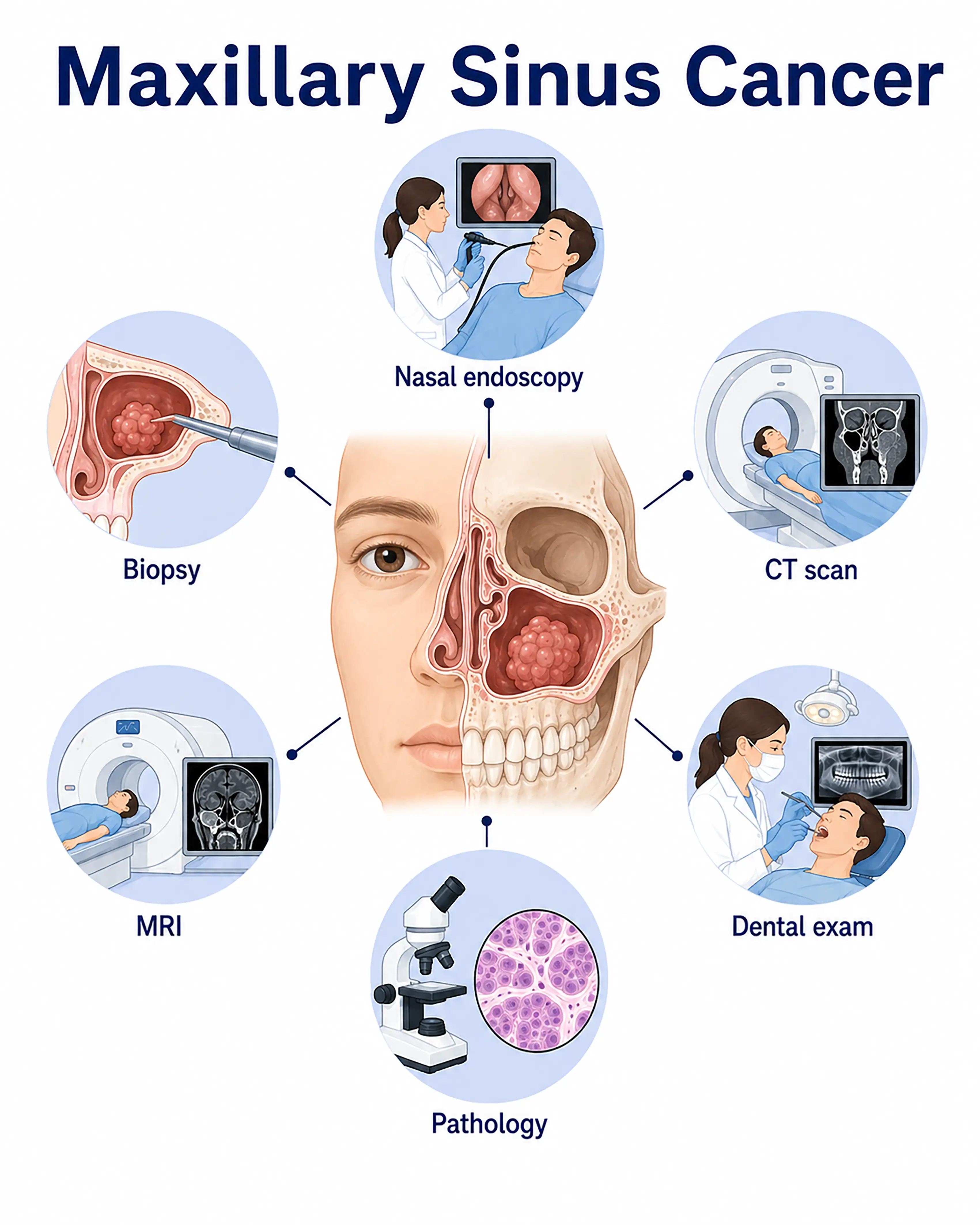
Maxillary sinus cancer diagnosis by stage
1.Early-stage maxillary sinus cancer diagnosis
The tumor is confined to the maxillary sinus mucosa, with symptoms resembling chronic sinusitis. Sinus CT shows soft-tissue opacity within the sinus with bone resorption or destruction. Endoscopic biopsy confirms the diagnosis.
2.Intermediate-stage maxillary sinus cancer diagnosis
The tumor destroys the sinus wall and invades the nasal cavity, orbit, or oral cavity. CT and MRI show extensive bone destruction and surrounding tissue involvement. Repeated biopsies are needed to establish the pathological type.
3.Advanced-stage maxillary sinus cancer diagnosis
The tumor extensively invades the skull base or facial skin, or spreads to distant sites. Whole-body PET-CT assesses metastatic extent, while needle biopsy or surgical exploration obtains sufficient tissue samples.
Without timely diagnosis, maxillary sinus cancer extensively destroys facial bones and invades important structures such as the orbit and skull base, making surgery extremely difficult and prognosis poor.
When to Seek Medical Attention
Early signs of maxillary sinus cancer include persistent unilateral nasal obstruction, facial swelling, toothache, and nosebleeds. Consult a doctor through "Chat with a Doctor" for early detection. During an online consultation, the doctor will ask about your symptoms and occupational exposure history and guide you toward sinus CT.
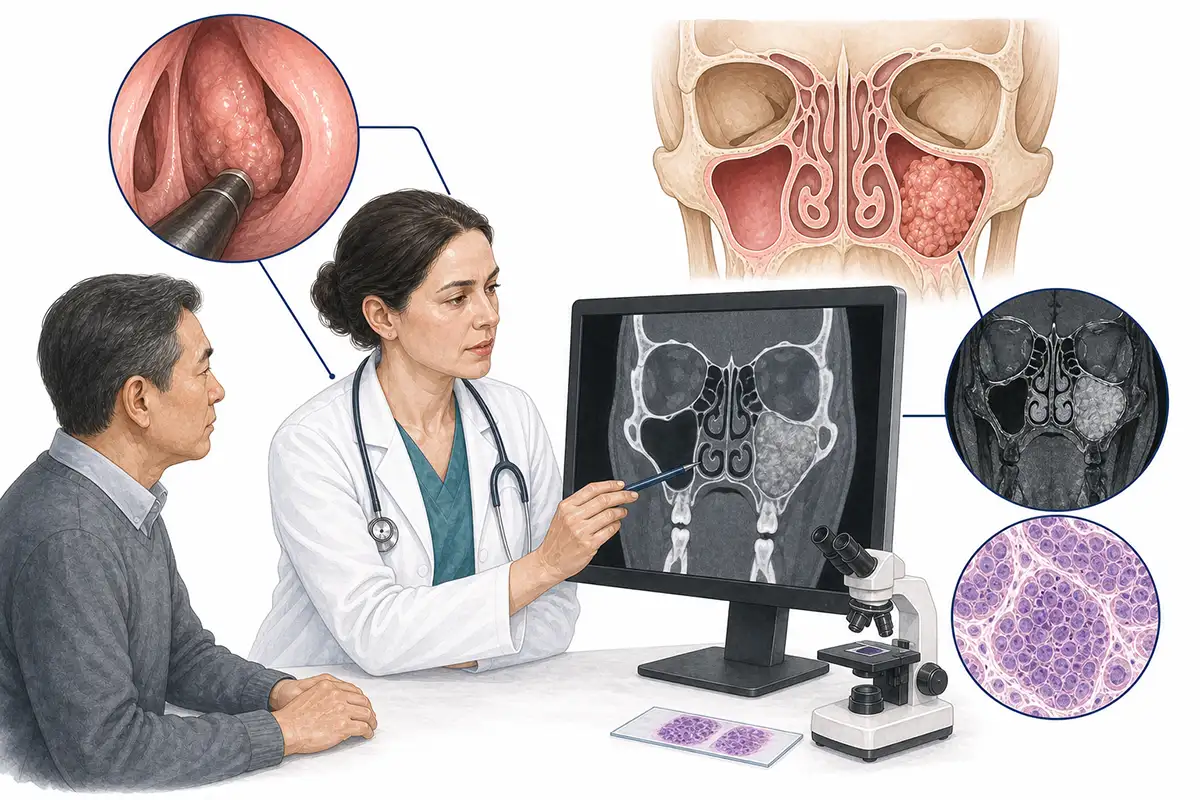
Basis for maxillary sinus cancer diagnosis
1.Sinus CT scan
Contrast-enhanced CT is the preferred imaging examination, showing tumor extent within the maxillary sinus, bone destruction, and invasion of surrounding structures to assess surgical feasibility.
2.Sinus MRI
MRI provides high soft-tissue resolution, clearly showing the tumor's relationship to surrounding muscles, blood vessels, and nerves and distinguishing tumor from inflammatory secretions.
3.Nasal endoscopy
Nasal endoscopy directly examines the nasal cavity and middle meatus, identifying tumor tissue protruding into the nasal cavity through the natural sinus opening and allowing endoscopy-guided biopsy.
4.Histopathological biopsy
Lesion tissue is obtained under nasal endoscopic or imaging guidance for pathological examination, the diagnostic gold standard. Squamous cell carcinoma is the most common pathological type.
5.Maxillary sinus needle biopsy
Maxillary sinus puncture obtains tissue from within the sinus for cytological or pathological examination and is suitable for lesions inaccessible by endoscopy.
6.Oral examination and assessment
Examine for loose upper teeth and tumor invasion of the gums and hard palate to assess the extent of downward tumor spread.
7.Eye examination and assessment
Assess eye movements, vision, and compression of orbital contents to determine the extent of upward orbital invasion.
8.Whole-body PET-CT assessment
Whole-body PET-CT assesses cervical lymph node and distant organ metastases, establishes tumor stage, and guides a comprehensive treatment plan.
9.Fine-needle aspiration biopsy
Ultrasound-guided fine-needle aspiration cytology of enlarged cervical lymph nodes assesses lymph node metastasis.
Daily Self-Check
Persistent unilateral nasal obstruction, facial swelling, toothache, or nosebleeds warrant prompt medical care. People with prolonged woodworking or nickel/chromium exposure should have regular sinus examinations.
Conclusion
Early symptoms of maxillary sinus cancer resemble sinusitis and are easily overlooked. Sinus CT and MRI are the main imaging assessment methods, and nasal endoscopic biopsy is the diagnostic gold standard.


