


Gliomas are tumors formed by the malignant proliferation of glial cells within the brain.Glial cells—including astrocytes, oligodendrocytes, and ependymal cells—serve to support and protect neurons.
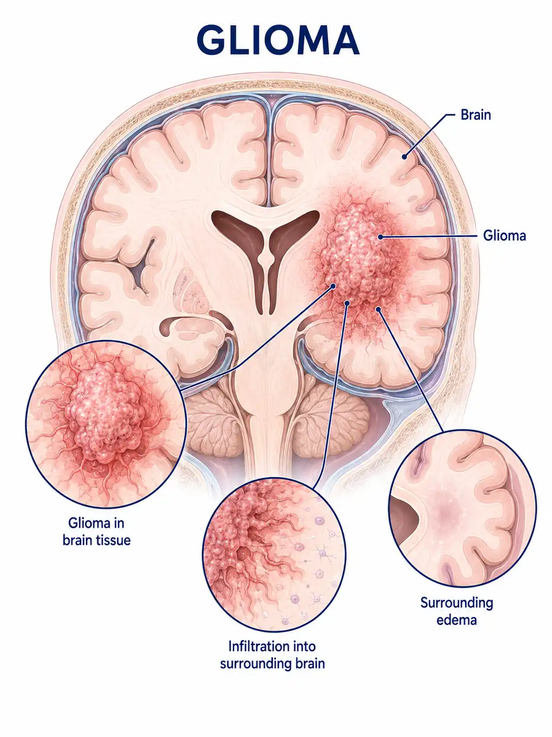
Global overview of gliomas
Gliomas account for a significant proportion of all primary brain tumors and can occur at any age, though certain types are more common in children.High-grade gliomas are difficult to treat and pose a major challenge in neurosurgery.
Consult a doctor regarding cancer-related issues
Individuals experiencing symptoms such as persistent headache, nausea and vomiting, seizures, limb weakness, or speech impairment should promptly consult a neurosurgeon or a neuro-oncologist.
Main types of gliomas
1. Astrocytoma
Originates in astrocytes; classified by the WHO into grades I–IV. Grade I tumors grow slowly and have a better prognosis, whereas Grade IV (glioblastoma) is the most malignant.
2. Oligodendroglioma
Originates in oligodendrocytes; often associated with 1p/19q co-deletion. Relatively sensitive to radiotherapy and chemotherapy; prognosis is comparatively favorable.
3. Types of ependymoma
Originates in ependymal cells; commonly occurs in the ventricular system. Can arise intracranially or in the spinal cord; in children, it is frequently found in the posterior cranial fossa.
4. Glioblastoma
It is the most malignant type of glioma, characterized by rapid growth, high invasiveness, and a tendency to recur; it is difficult to treat and carries a poor prognosis.
5. Mixed-type glioma
The tumor contains both astrocytic and oligodendroglial components; its biological behavior and response to treatment fall between those of the two cell types.
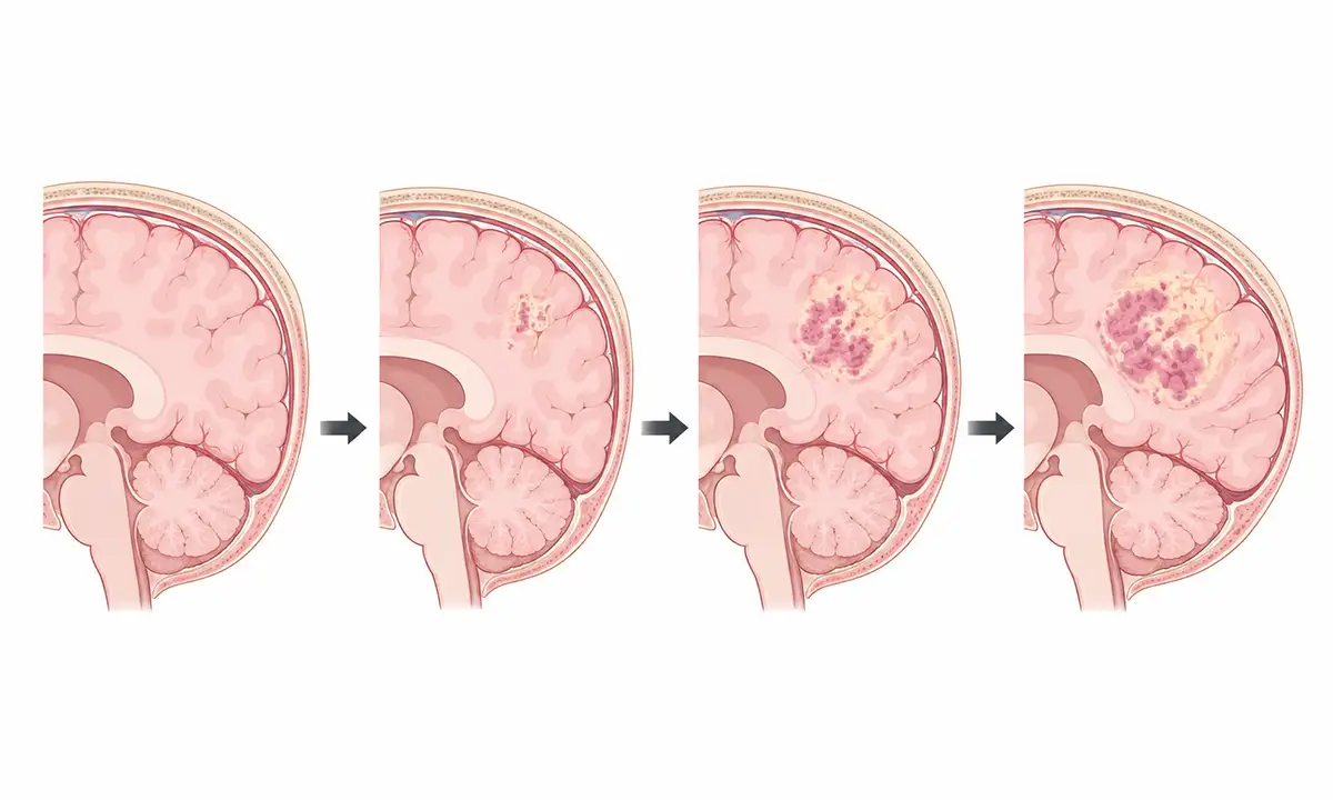
Typical symptoms and signs of gliomas
1. Headache and vomiting
A headache that gradually worsens upon waking, accompanied by nausea and vomiting, is a typical manifestation of increased intracranial pressure; the headache intensifies with coughing or straining.
2. Epileptic seizure
Seizures occurring without an obvious precipitating factor—especially a first-time seizure in an adult—warrant high vigilance, as they may be the initial symptom of a cerebral glioma.
3. Limb weakness
Gradual onset of weakness, numbness, or impaired mobility in a limb, associated with tumor compression of motor areas or nerve tracts.
4. Speech disorder
Difficulty with speech expression or comprehension, as well as difficulty finding words, may occur if the tumor affects the language centers of the brain.
5. Changes in vision
Decreased vision, visual field defects, or double vision (diplopia) may occur due to the tumor compressing the visual pathways or causing papilledema (swelling of the optic disc) resulting from increased intracranial pressure.
6. Cognitive changes
Memory loss, inability to concentrate, personality changes, and emotional disturbances may occur if the tumor affects the frontal or temporal lobes.
What are the diagnostic methods for glioma
1. Neurological physical examination
A physician assesses neurological deficits and localizes the site of the lesion by thoroughly examining vision, hearing, muscle strength, sensation, reflexes, and coordination.
2. Cranial MRI scan
MRI is the preferred method for diagnosing cerebral glioma; it reveals tumor location, size, morphology, and surrounding edema, while contrast-enhanced scanning aids in determining the tumor grade.
3. Pathological biopsy
Obtains tumor tissue via surgical resection or stereotactic biopsy; pathological examination serves as the gold standard for diagnosis and grading, accompanied by molecular testing.
4. Molecular testing
Detecting molecular markers such as IDH mutations, MGMT promoter methylation, and 1p/19q co-deletion provides important guidance for prognostic assessment and treatment selection.
5. Functional magnetic resonance imaging
fMRI can localize language and motor functional areas, aiding in the formulation of surgical plans to protect critical functional regions while resecting tumors.
6. Cerebrospinal fluid examination
Lumbar puncture is performed to obtain CSF for cytological examination, helping to rule out tumor dissemination via the cerebrospinal fluid.
What are the treatment methods for gliomas
1. Surgical treatment
Maximal safe resection is the primary treatment modality for gliomas, aiming to remove as much of the tumor as possible while preserving neurological function.
2. Radiation therapy
Postoperative adjuvant radiotherapy can reduce local recurrence rates; intensity-modulated radiotherapy and stereotactic radiotherapy allow for precise irradiation of the tumor while sparing surrounding normal brain tissue.
3. Chemotherapy
Temozolomide is a commonly used oral chemotherapy drug; its use concurrently with and as an adjuvant to radiotherapy can prolong patient survival.
4. Electric field therapy
Tumor-treating fields (TTFields) function by interfering with tumor cell mitosis; when combined with chemoradiotherapy, they can be used for newly diagnosed glioblastoma.
5. Targeted therapy
Anti-angiogenic drugs, such as bevacizumab, can alleviate cerebral edema and associated symptoms; they are used for recurrent or progressive gliomas.
6. Immunotherapy
Immune checkpoint inhibitors and tumor vaccines are being actively explored for glioma, with some clinical trials showing promise.
7. Symptomatic and supportive care
Antiepileptic drugs control seizures, glucocorticoids reduce cerebral edema to alleviate symptoms, and nutritional support improves the patient's general condition.
8. Rehabilitation follow-up
Postoperative follow-up includes regular MRI to monitor for recurrence; functional rehabilitation training helps improve neurological deficits, while psychological support enhances quality of life.
Conclusion
Gliomas are the most common primary malignant tumors of the central nervous system, with treatment primarily consisting of surgery combined with radiotherapy and chemotherapy.Early detection, maximal safe resection, and comprehensive treatment help extend patient survival and improve quality of life.


