


Gliomas account for approximately 80% of all malignant brain tumors.The WHO classifies gliomas into grades I through IV; Grade IV glioblastoma is the most common type and carries the poorest prognosis.The 2016 and 2021 WHO classification systems incorporated molecular markers into diagnostic criteria, with IDH mutation status serving as the most important indicator for molecular classification.
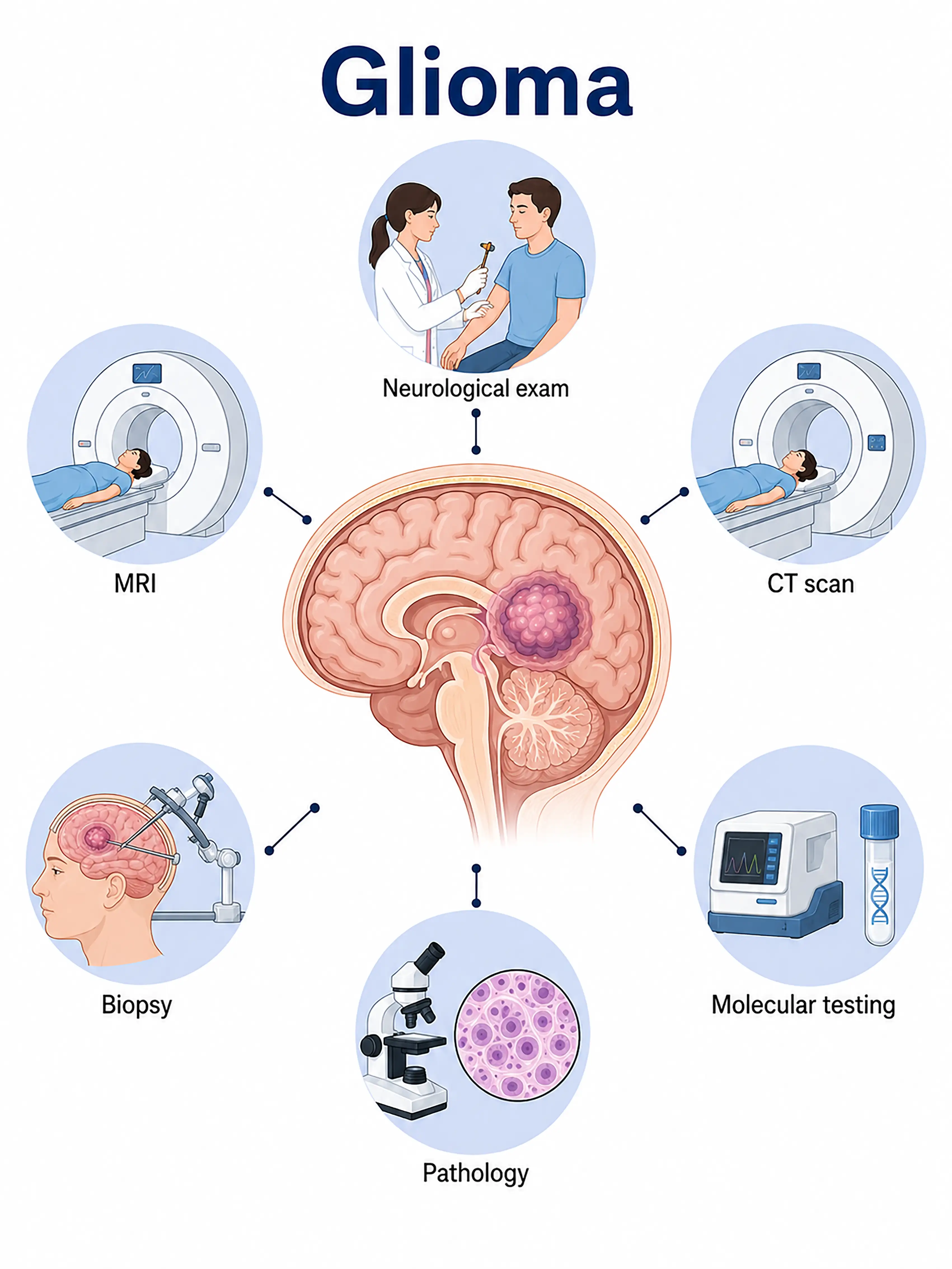
Diagnosis based on glioma stages
1.Early diagnosis of cerebral glioma
The tumor is small, slow-growing, and usually asymptomatic.An intracranial lesion with abnormal signal intensity is detected incidentally on MRI—hypointense on T1 and hyperintense on T2, with mild to moderate enhancement following contrast administration;biopsy is required for definitive diagnosis.
2.Diagnosis of intermediate-stage glioma
Headache, seizures, or focal neurological deficits.MRI shows an infiltrative tumor with surrounding edema and mass effect;obtain tissue via stereotactic or open-skull biopsy for pathological and molecular testing.
3.Diagnosis of advanced glioma
Severe elevation of intracranial pressure and extensive neurological impairment.MRI shows a large space-occupying lesion with midline shift and brain herniation; surgical specimen acquisition is performed to determine the WHO grade and molecular subtype, guiding the chemoradiotherapy regimen.
If not diagnosed promptly, gliomas—especially high-grade ones—can progress rapidly and extensively invade brain tissue;this leads to a missed window for optimal surgery and significantly shortens survival time.
When to Seek Medical Attention
Early signs of gliomas include persistent headaches, nausea and vomiting, seizures, and limb weakness.Please consult a doctor via the "Chat with Doctor" feature for early detection.During the consultation, the doctor will inquire about your symptoms and the progression of the condition, and advise you on undergoing a cranial MRI.
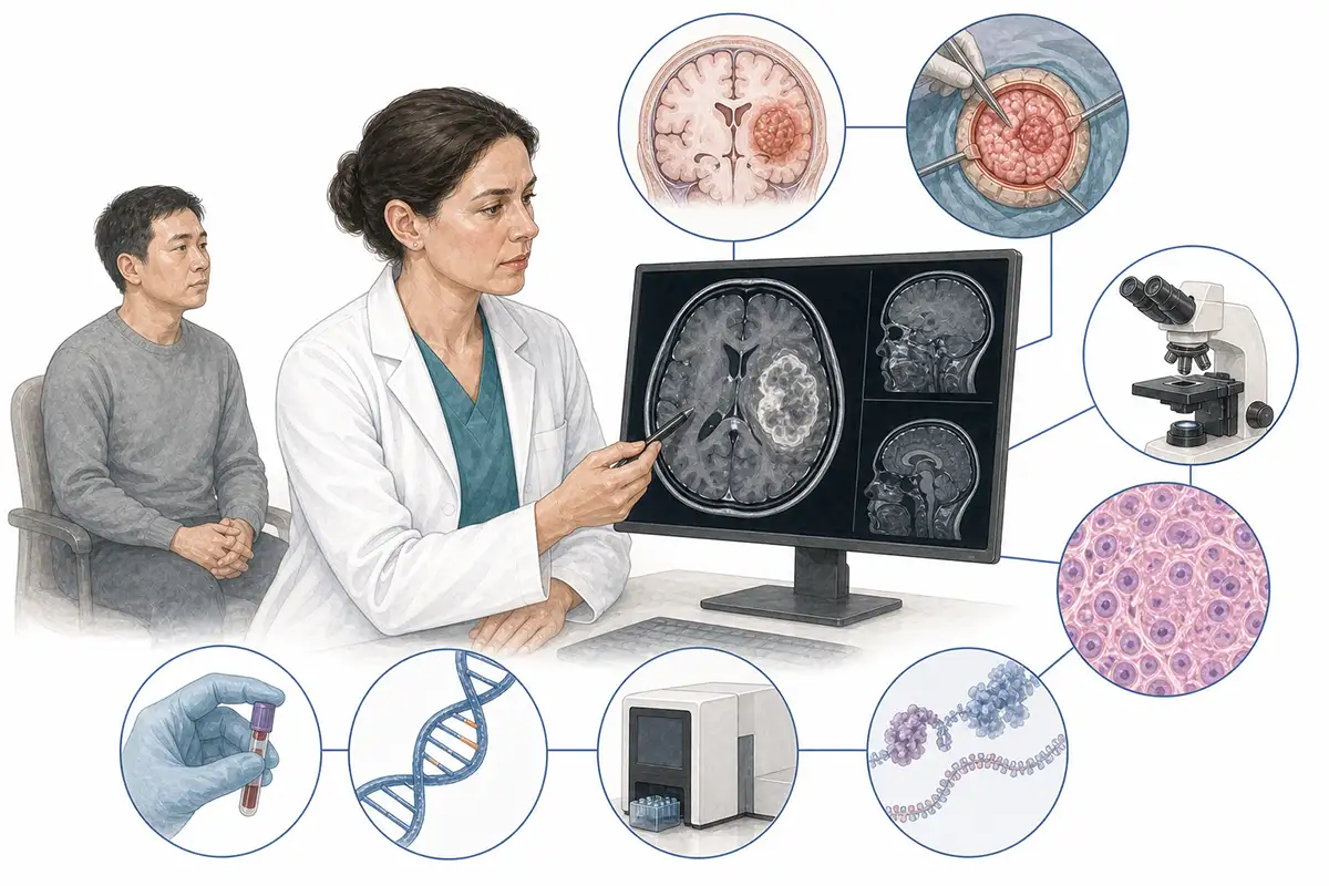
Basis for glioma diagnosis
1.Cranial MRI (plain + contrast-enhanced)
MRI is the preferred imaging modality for diagnosing cerebral glioma.Findings include low signal intensity on T1WI and high signal intensity on T2WI/FLAIR; enhancement patterns aid in grading the tumor.Multimodal MRI (DWI, PWI, MRS) assists in the assessment.
2.Histopathological biopsy
Pathological examination of tumor tissue obtained via stereotactic biopsy or craniotomy is the gold standard for the definitive diagnosis of glioma, allowing for the determination of histological type and WHO grade.
3.IDH gene testing
Determining IDH1/IDH2 gene mutation status is central to the molecular classification of gliomas; IDH-mutant tumors have a better prognosis than wild-type tumors, and this status serves as the primary stratification criterion in the 2021 WHO classification.
4. 1p/19q testing
Testing for the 1p/19q co-deletion status; its presence confirms a diagnosis of oligodendroglioma, which is sensitive to chemotherapy and serves as a molecular marker for a favorable prognosis.
5.MGMT promoter methylation
Detection of MGMT gene promoter methylation status; patients with positive methylation are more sensitive to temozolomide chemotherapy and have a better prognosis, making this a key marker for guiding treatment.
6.TERT promoter mutation testing
Detects *TERT* promoter mutations; when combined with *IDH* mutation status, this allows for the classification of gliomas into five molecular subgroups, serving as a key marker in the 2021 WHO classification.
7.CT scan
CT allows for rapid assessment of tumor size, calcification, and hemorrhage, as well as evaluation of brain herniation and hydrocephalus in emergency settings; it is superior to MRI for visualizing bone invasion.
8.PET-CT assessment
18F-FDG PET-CT distinguishes between low-grade and high-grade gliomas, assesses tumor metabolic activity, and aids in stereotactic biopsy localization and treatment efficacy evaluation.
9.Intraoperative rapid frozen section pathology
During surgery, rapid frozen-section analysis is performed on the obtained tissue to preliminarily determine the tumor's nature and grade, thereby guiding the extent of the resection.
Daily Self-Check
Seek medical attention promptly if unexplained persistent headache, seizures, limb weakness, or personality changes occur.Regular physical examinations help in the early detection of asymptomatic lesions.
Conclusion
Diagnosis of gliomas relies on MRI and pathological biopsy; molecular testing (e.g., IDH, 1p/19q, MGMT) forms the basis for modern classification and treatment decision-making.


