


Central nervous system tumors include primary brain tumors and spinal cord tumors; gliomas are the most common primary malignant brain tumors. The annual incidence rate of brain gliomas in my country is 5–8 per 100,000 people.The 2021 WHO classification incorporated molecular markers into diagnostic criteria, advancing the development of precision diagnosis and treatment.
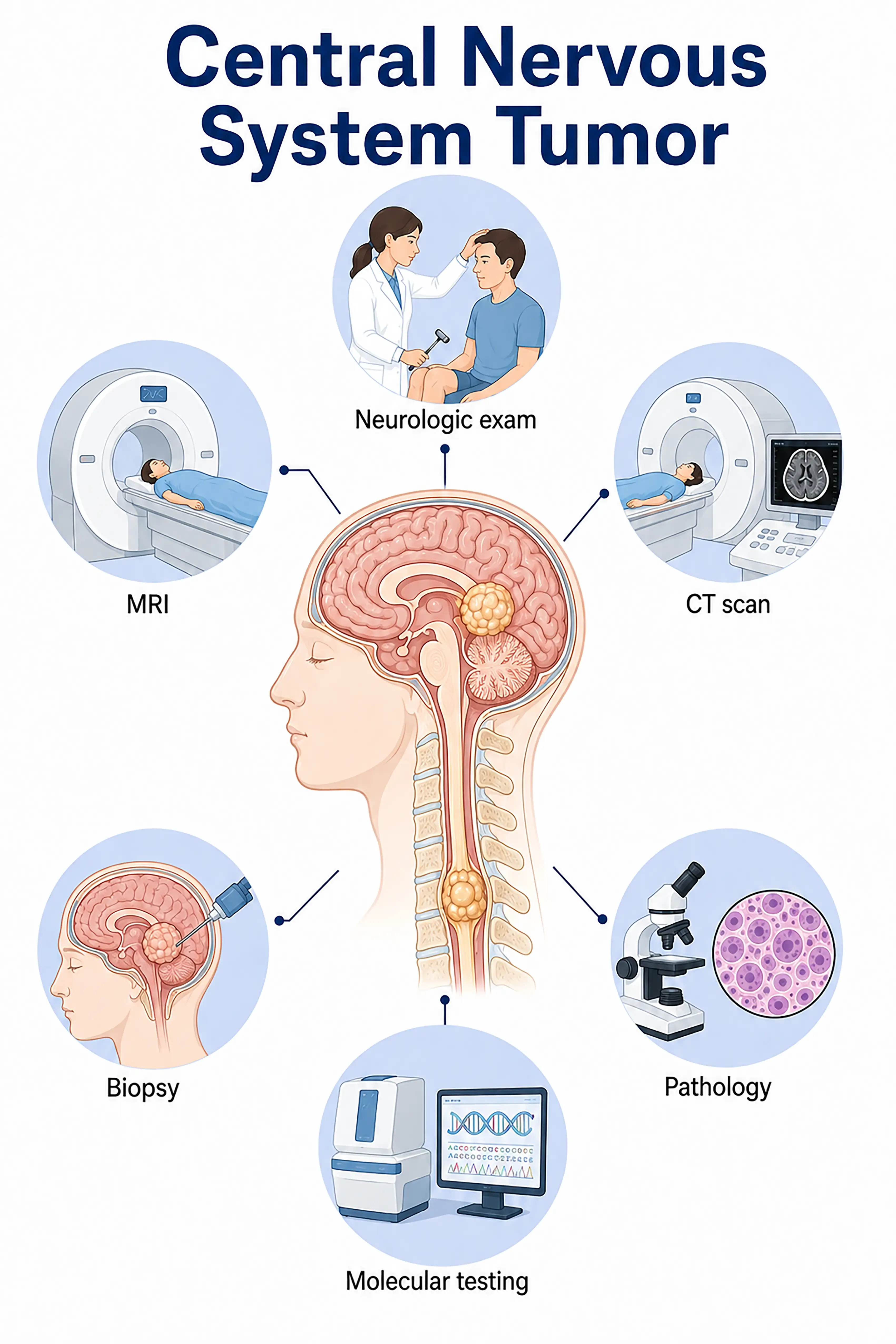
Diagnosis based on central nervous system tumor stages
1.Early diagnosis of central nervous system tumors
An intracranial or intraspinal lesion with abnormal signal intensity is incidentally detected on MRI.It appears hypointense on T1WI and hyperintense on T2WI, with variable enhancement (or lack thereof) following contrast administration; stereotactic or surgical biopsy is required for confirmation.
2.Diagnosis of intermediate-stage CNS tumors
Headache, seizures, or focal neurological deficits.MRI shows a tumor with surrounding edema and mass effect; obtain a specimen via surgery or biopsy to determine the pathological type and WHO grade.
3.Diagnosis of advanced central nervous system tumors
Severe elevation of intracranial pressure and extensive neurological impairment.MRI shows a large space-occupying lesion with midline shift and brain herniation; surgical decompression and specimen acquisition are performed, with molecular testing guiding the comprehensive treatment plan.
If not diagnosed promptly, CNS tumors—especially high-grade ones—will progress rapidly, compressing vital brain regions or the spinal cord and causing irreversible neurological damage.
When to Seek Medical Attention
Early signs of central nervous system tumors include persistent headaches, nausea and vomiting, seizures, and limb weakness.Consult a doctor via "Chat with a Doctor" for early detection.During the online consultation, the doctor will ask about your symptoms and their progression, and guide you through the process of getting an MRI scan.
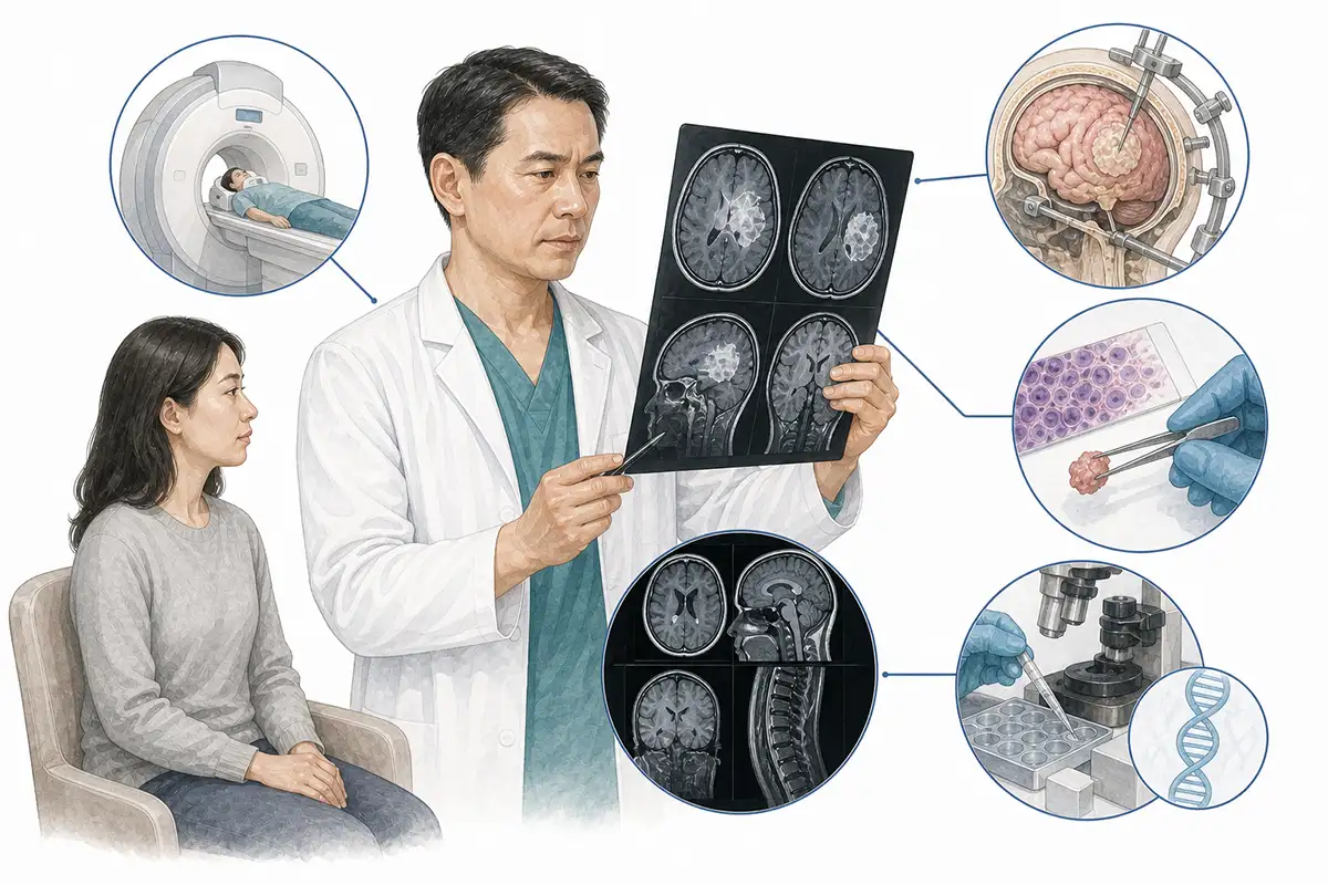
Diagnostic criteria for CNS tumors
1.Cranial/spinal MRI
MRI is the preferred imaging modality for diagnosing central nervous system tumors, clearly showing tumor location, size, morphology, and peritumoral edema; post-contrast enhancement characteristics help determine the nature of the tumor.
2.Multimodal MRI
Multimodal MRI techniques—including DWI, DTI, PWI, and MRS—assist in differentiating between neoplastic and non-neoplastic lesions and in assessing tumor malignancy and the extent of infiltration.
3.Stereotactic biopsy
Precise image-guided needle biopsy to obtain tumor tissue samples for pathological and molecular testing is a key method for diagnosing deep-seated tumors or those located in functional areas.
4.Craniotomy/Laminectomy
Surgical resection to obtain a specimen for integrated histopathological and molecular pathological diagnosis, while simultaneously reducing tumor burden, is a crucial diagnostic and therapeutic approach.
5.IDH gene testing
Determining IDH1/IDH2 gene mutation status serves as the primary stratification criterion in the 2021 WHO classification; IDH-mutant tumors have a better prognosis than wild-type tumors.
6.1p/19q testing
Testing for the 1p/19q co-deletion status; its presence confirms a diagnosis of oligodendroglioma, which is sensitive to chemotherapy and serves as a molecular marker for a favorable prognosis.
7.CT scan
CT allows for rapid assessment of tumor size, calcification, and hemorrhage, as well as evaluation of brain herniation and hydrocephalus in emergency settings; it is superior to MRI for visualizing bony structures.
8.PET-CT assessment
18F-FDG PET-CT can differentiate between low-grade and high-grade tumors, assess tumor metabolic activity, and assist in staging and treatment efficacy evaluation.
9Intraoperative neurophysiological monitoring
Real-time intraoperative monitoring of neurological function protects vital functional areas, improving both surgical safety and the rate of tumor resection.
Daily Self-Check
Seek medical attention promptly if unexplained persistent headache, seizures, limb weakness, or sensory abnormalities occur.Regular physical examinations help in the early detection of asymptomatic lesions.
Conclusion
Diagnosis of CNS tumors relies on MRI and pathological biopsy.Molecular testing (e.g., IDH, 1p/19q) forms the basis for modern classification and treatment decisions.


