


Buccal mucosal cancer is relatively common among oral cancers and is one of the high-incidence malignancies in Southeast Asia, closely associated with betel nut chewing. It usually affects men aged 40-60, and squamous cell carcinoma is the main pathological type.
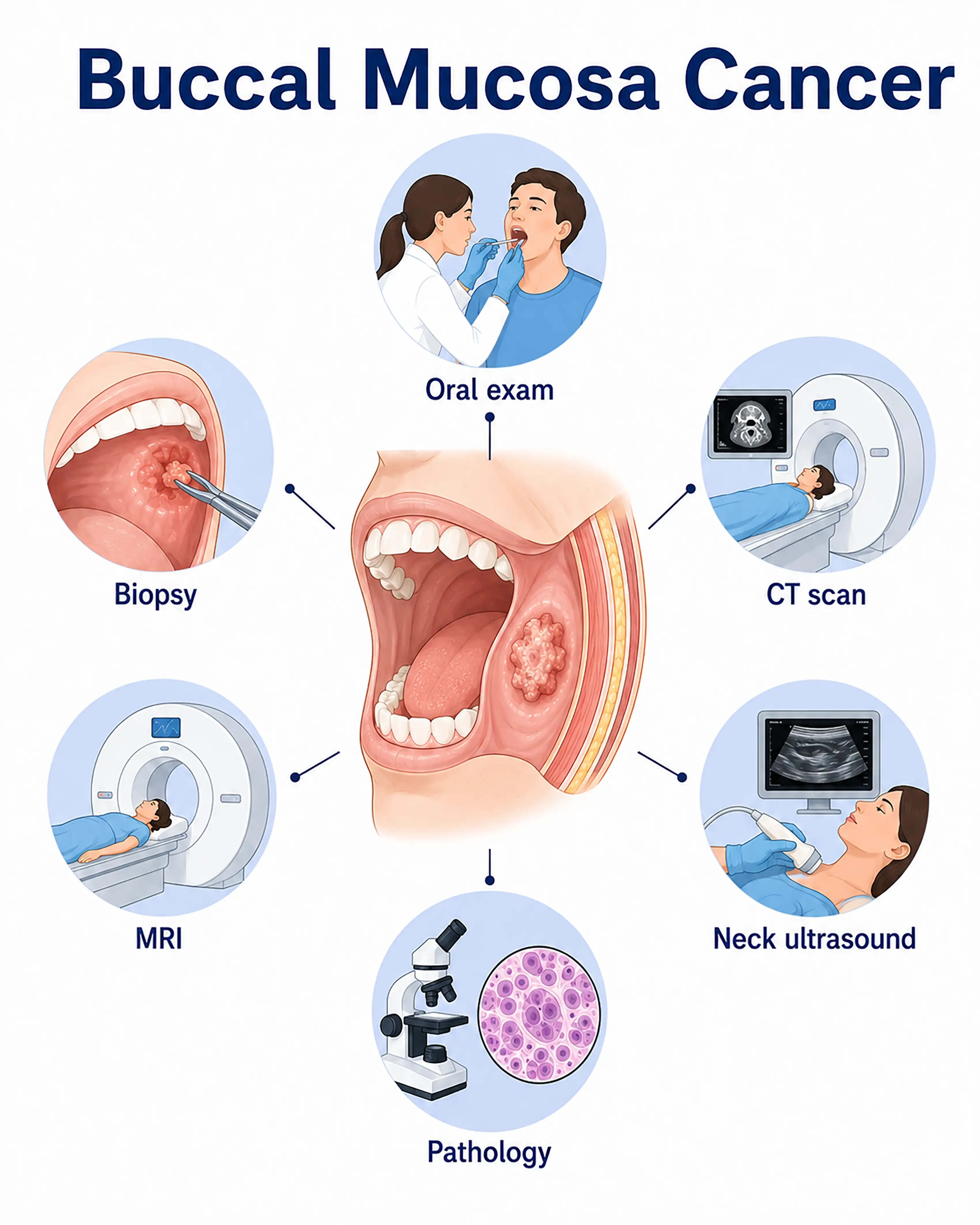
Buccal mucosal cancer diagnosis by stage
1.Early-stage buccal mucosal cancer diagnosis
The tumor is confined to the mucosal layer, presenting as white patches, red patches, or small ulcers. Oral examination and palpation reveal roughness or induration of the cheek mucosa, and biopsy confirms the diagnosis.
2.Intermediate-stage buccal mucosal cancer diagnosis
The tumor infiltrates deeper layers and forms an obvious lump or ulcer. CT or MRI assesses invasion extent and cervical lymph node metastasis, while biopsy determines pathological type and differentiation.
3.Advanced-stage buccal mucosal cancer diagnosis
The tumor invades the mandible or chewing muscles, or spreads to distant sites. Whole-body PET-CT assesses distant metastasis, surgical exploration establishes the stage, and pathology guides comprehensive treatment.
Without timely diagnosis, buccal mucosal cancer invades the mandible and chewing muscles, causing difficulty opening the mouth and eating. Cervical lymph node metastasis is common, and prognosis declines significantly.
When to Seek Medical Attention
Early signs of buccal mucosal cancer include white or red buccal patches, long-standing nonhealing ulcers, cheek lumps, and restricted mouth opening. Consult a doctor through "Chat with a Doctor" for early detection. During an online consultation, the doctor will ask about your oral hygiene habits and symptoms and guide you toward oral examination and biopsy.
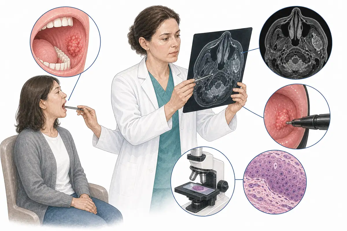
Basis for buccal mucosal cancer diagnosis
1.Clinical oral examination
Visual examination and palpation assess changes in cheek mucosal color, consistency, and shape, identifying white patches, red patches, ulcers, or lumps and evaluating lesion extent and mobility.
2.Histopathological biopsy
Pathological examination of suspicious tissue obtained by incisional biopsy is the gold standard for diagnosing buccal mucosal cancer and determines pathological type and differentiation.
3.Oral and maxillofacial CT
Contrast-enhanced CT assesses invasion of cheek soft tissue, the mandible, and surrounding structures and determines whether bone destruction is present.
4.Oral and maxillofacial MRI
MRI provides high soft-tissue resolution, assessing the tumor's relationship to chewing muscles, blood vessels, and nerves and distinguishing tumor from inflammation.
5.Cervical lymph node ultrasound
Assesses cervical lymph node metastasis and guides needle biopsy of suspicious nodes to establish tumor stage.
6.Whole-body PET-CT assessment
Whole-body PET-CT assesses distant metastasis and detects small metastatic lesions that conventional imaging may miss, enabling precise staging.
7.Exfoliative cytology
Cells scraped from the lesion surface are examined on a smear. Detection of cancer cells supports diagnosis, and the method is suitable for superficial lesions.
8.Narrow-band imaging endoscopy
Special light wavelengths enhance visualization of mucosal surface vessels, helping detect early precancerous lesions and define tumor boundaries.
9.HPV testing
Some buccal mucosal cancers are associated with HPV infection. HPV testing helps analyze the cause and assess prognosis.
Daily Self-Check
Examine the oral mucosa regularly and seek prompt care for white patches, red patches, or ulcers that do not heal within two weeks. Avoiding betel nut chewing, quitting smoking, and limiting alcohol are important preventive measures.
Conclusion
Early symptoms of buccal mucosal cancer are easily overlooked. Oral white patches or long-standing nonhealing ulcers warrant prompt medical evaluation. Quitting smoking, limiting alcohol, and avoiding betel nut chewing are important preventive measures.


