


Tongue cancer is the most common oral cancer, accounting for about 30%-40% of oral malignancies. Men are affected more often, usually at ages 40-60. Smoking, drinking, and HPV infection are major risk factors. It comprises oral tongue and tongue base cancer, with the former having a relatively better prognosis.
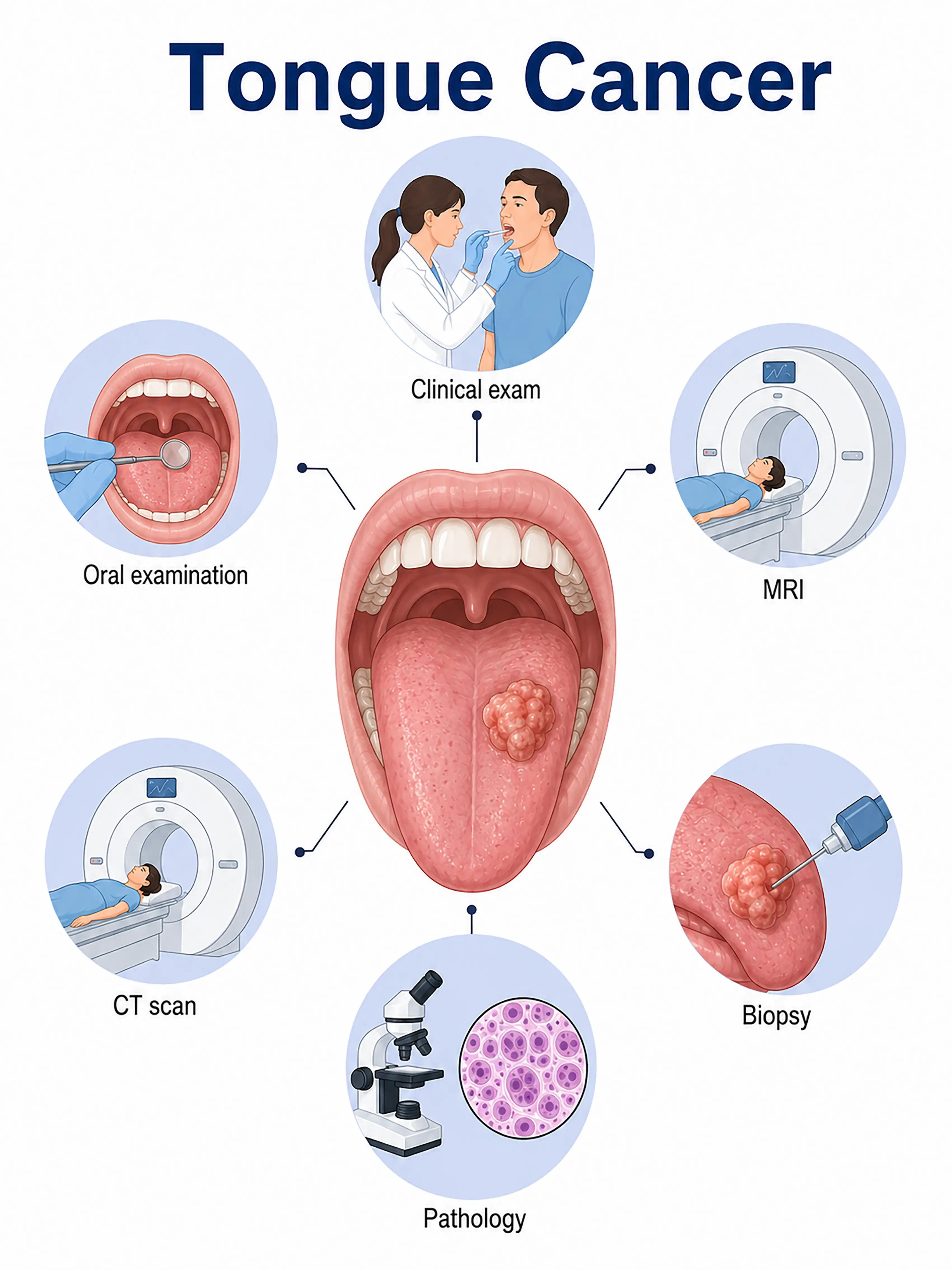
Tongue cancer diagnosis by stage
1.Early-stage tongue cancer diagnosis
The tumor is confined to the tongue mucosa, presenting as a small ulcer or white patch on the tongue border. Oral examination and palpation reveal induration. Incisional biopsy at the tumor edge provides pathological confirmation.
2.Intermediate-stage tongue cancer diagnosis
The tumor enlarges and infiltrates tongue muscles, causing marked pain and restricted movement. Contrast-enhanced CT and MRI assess tumor extent and cervical lymph node metastasis, while biopsy determines the pathological type.
3.Advanced-stage tongue cancer diagnosis
The tumor invades the floor of the mouth or mandible, or spreads to distant sites. Whole-body PET-CT assesses distant metastasis, surgical exploration establishes the stage, and pathology guides comprehensive treatment.
Without timely diagnosis, tongue cancer extensively invades tongue muscles and surrounding tissues, severely impairing speech and swallowing. Cervical lymph node metastasis is common, and prognosis declines significantly.
When to Seek Medical Attention
Early signs of tongue cancer include a tongue ulcer lasting more than two weeks, a tongue lump, worsening pain, and restricted movement. Consult a doctor through "Chat with a Doctor" for early detection. During an online consultation, the doctor will ask about your oral hygiene habits and symptoms and guide you toward oral examination and biopsy.
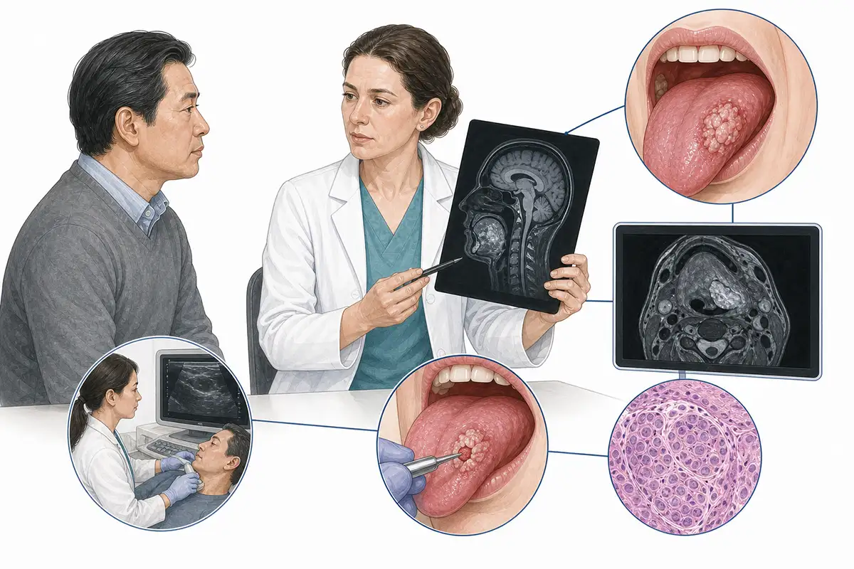
Basis for tongue cancer diagnosis
1Clinical oral examination
Visual examination and palpation assess color, consistency, and shape changes in the tongue mucosa, lateral borders, and underside, evaluating ulcer or lump location, size, and mobility.
2.Histopathological biopsy
Incisional biopsy at the tumor edge obtains tissue for pathological examination, the diagnostic gold standard. Definitive surgery should await a confirmed pathological diagnosis.
3.Neck ultrasound
High-frequency ultrasound assesses cervical lymph node metastasis, particularly the upper deep cervical nodes, and guides biopsy of suspicious nodes.
4.Head and neck CT scan
Contrast-enhanced CT assesses tumor extent, proximity to important structures, and cervical lymph node metastasis and is commonly used for clinical staging and treatment planning.
5.Head and neck MRI
MRI has distinct advantages for soft-tissue lesions and better assesses infiltration between tongue muscle fibers, complementing CT.
6.Panoramic X-ray examination
Panoramic radiographs assess mandibular invasion and help determine the extent of downward tumor spread.
7.Chest CT scan
As the preferred examination for lung metastasis screening, chest CT should be added when lung metastasis is suspected to establish the diagnosis.
8.Whole-body PET-CT assessment
Whole-body PET-CT assesses distant metastasis and synchronous second primary cancers and is recommended when residual or recurrent tumor is suspected.
9.HPV testing
Some tongue base cancers are associated with HPV infection. HPV testing supports etiological analysis, prognosis assessment, and treatment decisions.
Daily Self-Check
Seek prompt care for tongue ulcers persisting beyond two weeks, tongue lumps, or worsening pain. Have regular oral examinations, quit smoking, limit alcohol, and avoid betel nuts.
Conclusion
Tongue cancer is the most common oral cancer. Early diagnosis is crucial to preserving tongue function and improving survival. Oral examination combined with pathological biopsy is key to confirmation.


