


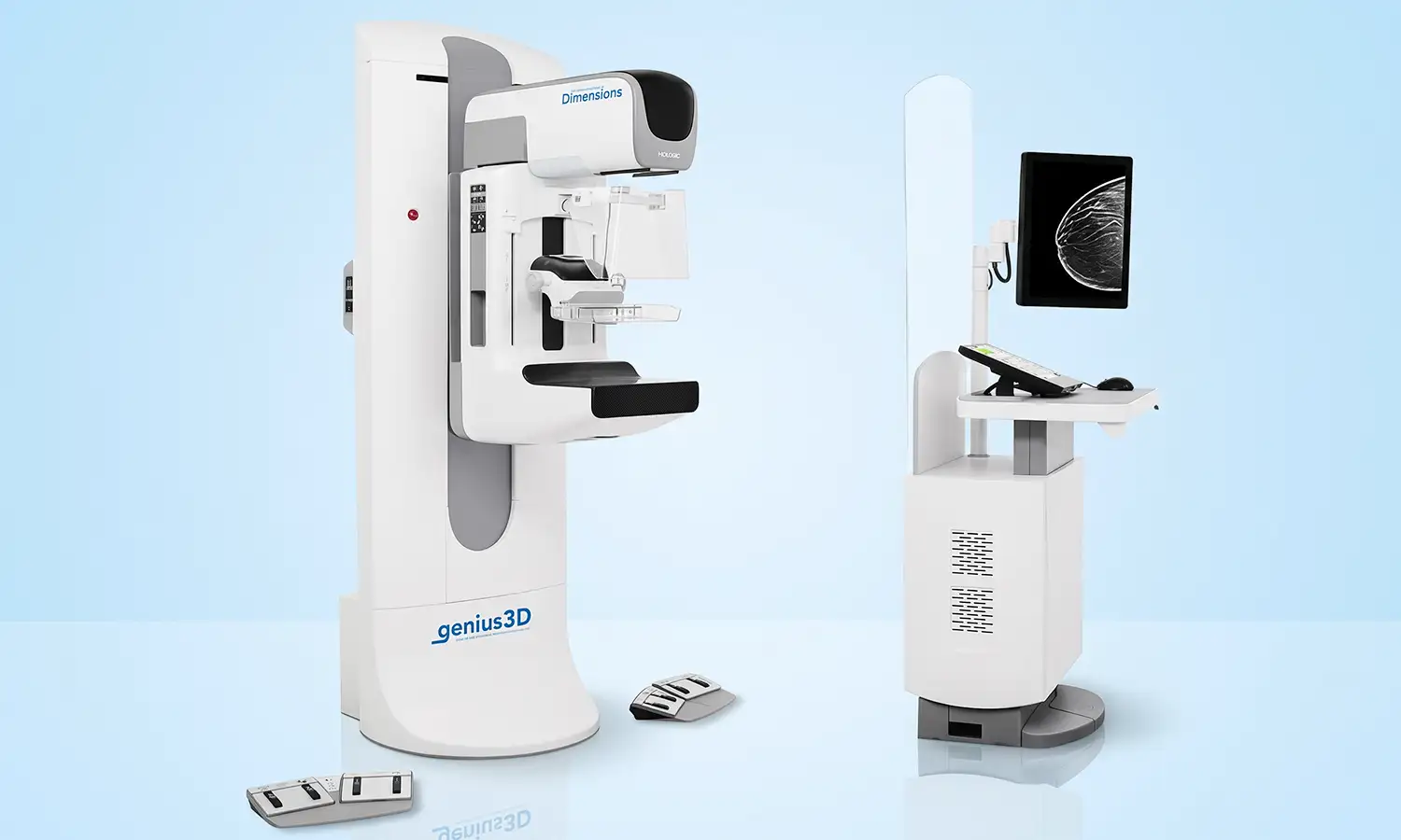
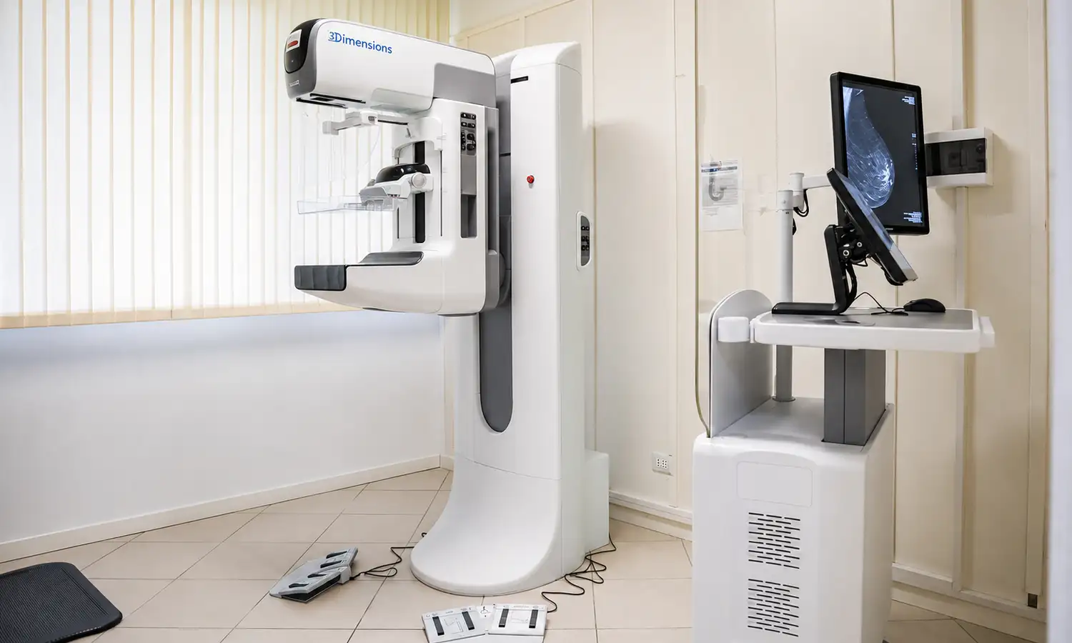
The Hologic 3Dimensions™ Digital Breast Tomosynthesis System (USA) is a world-leading diagnostic imaging system for women's health, designed for the early screening, diagnosis, and precise localization of breast cancer.The product line includes the Selenia Dimensions series and the next-generation 3Dimensions Mammography System. It supports capabilities such as 2D digital mammography, 3D tomosynthesis, intelligent image processing, and stereotactic biopsy, providing high-quality imaging for breast cancer screening, clinical diagnosis, and preoperative assessment.By combining advanced digital mammography with Digital Breast Tomosynthesis (DBT) technology—utilizing multi-angle image acquisition and 3D reconstruction—the system enhances the visualization of breast tissue details, enabling physicians to detect tiny, abnormal lesions that might otherwise be obscured by overlapping tissue in conventional 2D mammography.Featuring a high-resolution detector, advanced image reconstruction algorithms, and a patient-friendly design, this equipment enhances lesion detection capabilities while optimizing the patient's examination experience;suitable for breast disease screening and precision diagnosis, it is a key piece of equipment for modern breast imaging centers.
Core Technologies
What is the HOLOGIC digital mammography system?
The HOLOGIC digital mammography system is specialized imaging equipment that uses low-dose X-rays to capture images of breast tissue and employs digital image processing and 3D reconstruction technologies to assist in the diagnosis of breast diseases.The system clearly displays internal structural changes—including microcalcifications, abnormal tissue thickening, and imaging signs associated with early-stage tumors—providing physicians with a more comprehensive basis for diagnosis.A key technical feature of HOLOGIC equipment is 3D breast tomosynthesis.This technology acquires breast images from multiple angles and reconstructs them into 3D images similar to tomographic scans, allowing physicians to examine tissue structures at various depths. This effectively mitigates diagnostic challenges caused by tissue overlap in traditional 2D mammography and enhances the ability to identify lesions within complex breast tissue environments.
1. 3D breast tomosynthesis technology
HOLOGIC equipment utilizes advanced 3D breast tomosynthesis technology; by continuously acquiring breast image data from various angles and employing high-performance computing algorithms for 3D reconstruction, it allows breast tissue to be visualized across multiple planes.This technology mitigates the impact of tissue overlap on image interpretation, enabling physicians to clearly observe abnormal areas that might otherwise be obscured by dense breast tissue.Compared to traditional 2D mammography, 3D tomosynthesis provides richer spatial information and enhances the detection of early-stage breast cancer and minute lesions, making it particularly suitable for patients with high breast density or complex imaging characteristics.
2. High-definition digital detection and image processing technology
HOLOGIC mammography equipment utilizes high-performance digital detectors and advanced image processing technology to acquire high-resolution breast images and enhance the visualization of minute structures and abnormal areas.By optimizing image acquisition parameters and reconstruction algorithms, the system controls radiation dosage while maintaining diagnostic image quality, allowing physicians to obtain clearer and more stable imaging data.High-definition digital imaging technology assists radiologists in accurately analyzing changes in breast tissue, thereby improving the efficiency of screening and diagnosis.
3. Intelligent image analysis and computer-aided diagnosis technology
The HOLOGIC system integrates intelligent image analysis software to assist in processing breast images, thereby enhancing the efficiency of image interpretation and diagnostic consistency for physicians.Through digital image management and intelligent analysis capabilities, the equipment enables physicians to rapidly pinpoint suspicious areas and make comprehensive assessments by incorporating patient clinical data, providing an imaging basis for subsequent examinations and treatment planning.This technology helps streamline breast screening workflows, allowing medical institutions to deliver more standardized and consistent early breast cancer screening services.
4. Stereotactic biopsy technique
HOLOGIC mammography equipment supports stereotactic biopsy capabilities, enabling precise localization of abnormal areas identified in images to guide physicians in tissue sampling.This technology is particularly suitable for locating microcalcifications, non-palpable breast abnormalities, and lesions highly suspected based on imaging.Precise image guidance improves biopsy accuracy, provides reliable tissue samples for pathological diagnosis, and ensures an effective link between initial imaging detection and precise diagnosis.
Scope of Indications
HOLOGIC mammography equipment is primarily used for the screening, diagnosis, and precise localization of breast diseases.
1. Areas for examination/adjunctive diagnosis
Application Field Primary Application
Breast cancer screening Early screening for breast cancer in women and regular check-ups for high-risk groups
Diagnosis of breast diseases Assessment of breast masses, microcalcifications, and structural abnormalities
Preoperative imaging assessment: determining the extent of the lesion and assisting in the formulation of the treatment plan.
Biopsy localization: Precise image-guided localization of suspected lesions
2. Target patient population
• Individuals requiring breast cancer screening
For women eligible for breast cancer screening, HOLOGIC equipment employs high-definition digital imaging and 3D tomosynthesis technology to detect early-stage abnormalities, enhancing the ability to identify the disease at an early stage.
• Patients with abnormal breast imaging findings
For patients with abnormal findings on breast palpation, suspicious areas detected via ultrasound, or abnormalities in previous imaging, this device provides detailed imaging information to assist physicians in further characterizing the lesion.
• High-risk individuals and those with dense breast tissue
For individuals at high risk for breast cancer or those with dense breast tissue, 3D tomosynthesis technology minimizes the impact of tissue overlap, thereby improving the ability to detect abnormalities within complex breast structures.
Core Strengths
Comparison Dimension Conventional 2D Mammography Hologic Mammography Equipment
Imaging mode 2D planar imaging Supports 3D tomographic imaging
Tissue visualization; susceptible to tissue overlap; multi-slice imaging enhances visualization capabilities
Lesion detection Limited ability to identify minute abnormalities Enhanced capability to detect early-stage abnormalities
Image Quality Dependent on equipment performance High-definition digital detection and intelligent processing
Clinical Use: Primarily for screening; integrated application for screening, diagnosis, and localization
Patient experience Limited comfort during examination Optimized compression design; improved examination experience
Conventional 2D Mammography
Conventional 2D mammography primarily visualizes breast tissue structures through single-plane images; however, the overlapping of internal breast tissues can obscure minute abnormalities with normal tissue, thereby compromising the visibility of lesions.During early screening for breast disease, the dimensionality of the imaging data is relatively limited, presenting challenges for the further diagnosis and precise localization of complex cases.
HOLOGIC Digital Breast X-ray Equipment
HOLOGIC breast X-ray equipment employs advanced digital mammography technology and supports 3D tomosynthesis; by reconstructing images across multiple layers, it minimizes the effects of tissue overlap and improves the visualization of breast structures and abnormal lesions.Compared to traditional 2D mammography, the system utilizes high-definition digital detectors and intelligent image processing to enhance image quality, facilitating the detection of subtle, early-stage abnormalities.Additionally, the equipment supports various applications—including breast disease screening, clinical diagnosis, and lesion localization—and features an optimized breast compression design that ensures high image quality while enhancing patient comfort during the examination, thereby providing essential imaging support for the early detection and precise diagnosis and treatment of breast diseases.
FAQ
1. What is the difference between 3D breast tomosynthesis and standard 2D mammography?
3D breast tomosynthesis acquires low-dose projection data from multiple angles and reconstructs them into thin-slice images, helping to reduce the impact of overlapping breast tissue on visualization.The decision to use 2D, 3D, or combined imaging should be made by a physician based on screening or diagnostic needs.
2. Is breast compression required for 3D imaging exams?
Yes, it is necessary. Appropriate compression helps stabilize the breast, spread out the tissue, and reduce motion artifacts, while also helping to control the radiation dose.Compression typically lasts only a short time; if you experience significant pain, please inform the technician immediately.
3. Is the radiation dose high for 3D breast imaging?
3D breast imaging uses X-rays; the specific radiation dose depends on the selected imaging mode, breast thickness, and whether 2D images are acquired simultaneously.Medical institutions select appropriate protocols based on the purpose of the examination and adhere to the principle of dose optimization.
4. Do individuals with dense breast tissue have a greater need for 3D imaging?
3D tomographic images may help reduce tissue overlap; however, determining whether patients with dense breasts require 3D imaging—or a combination of ultrasound and MRI—requires an assessment of age, symptoms, family history, and overall risk, rather than relying solely on breast density.
5. How do I schedule a HOLOGIC 3Dimensions 3D mammography exam?
Before making an appointment, please inform the breast specialist or radiology department of your screening or diagnostic needs, and disclose any pregnancy, potential pregnancy, or breast implants.Please bring previous breast imaging records and reports; if you have implants, please specify the type, date of insertion, and any concerns regarding pain or rupture so that appropriate examination and positioning protocols can be arranged.