


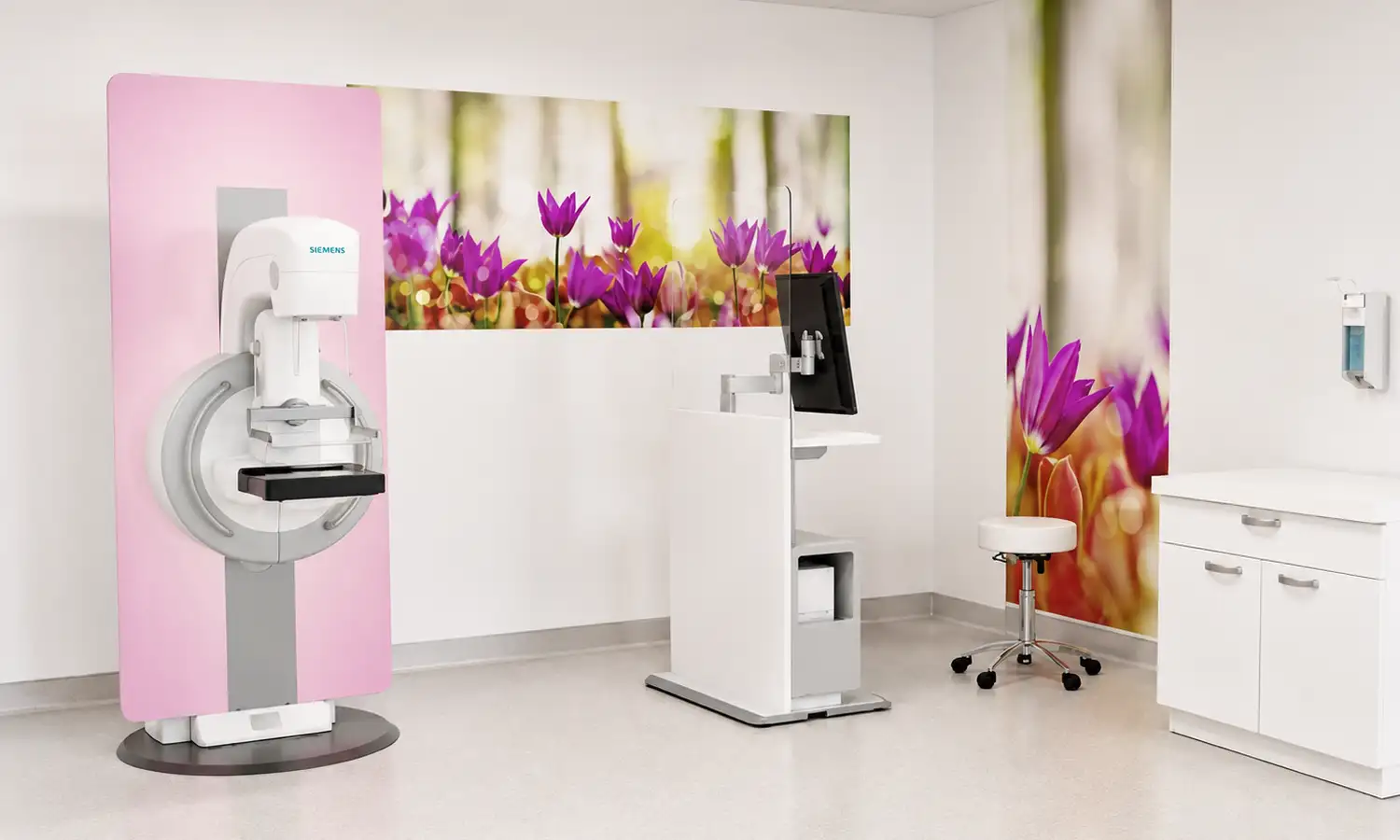
The Siemens MAMMOMAT digital mammography system, developed by Siemens Healthineers, is specialized equipment designed for breast imaging.The system comprises an X-ray generator, a digital flat-panel detector, a mammography platform, an image acquisition and processing workstation, an image display system, and auxiliary positioning devices, enabling high-quality image acquisition and intelligent image analysis.By optimizing breast imaging technology, the system ensures image clarity while minimizing unnecessary radiation exposure, providing physicians with clear and reliable information on breast tissue structures; it is particularly suitable for early breast cancer screening and the detection of abnormalities such as microcalcifications and masses.
Core Technologies
What is the MAMMOMAT digital mammography system?
MAMMOMAT is a digital X-ray imaging system dedicated to breast examinations; it captures images of breast tissue using high-resolution digital detectors and employs advanced image processing to enhance the visualization of lesions.Compared to traditional mammography, digital mammography yields clearer tissue details, allowing physicians to more accurately observe structural changes within the breast.In addition to standard 2D mammography, the system integrates 3D tomosynthesis technology to capture breast tissue data from multiple angles, thereby minimizing the impact of tissue overlap on lesion visibility and improving the detection of abnormalities within complex breast structures.
1. High-definition digital breast imaging technology
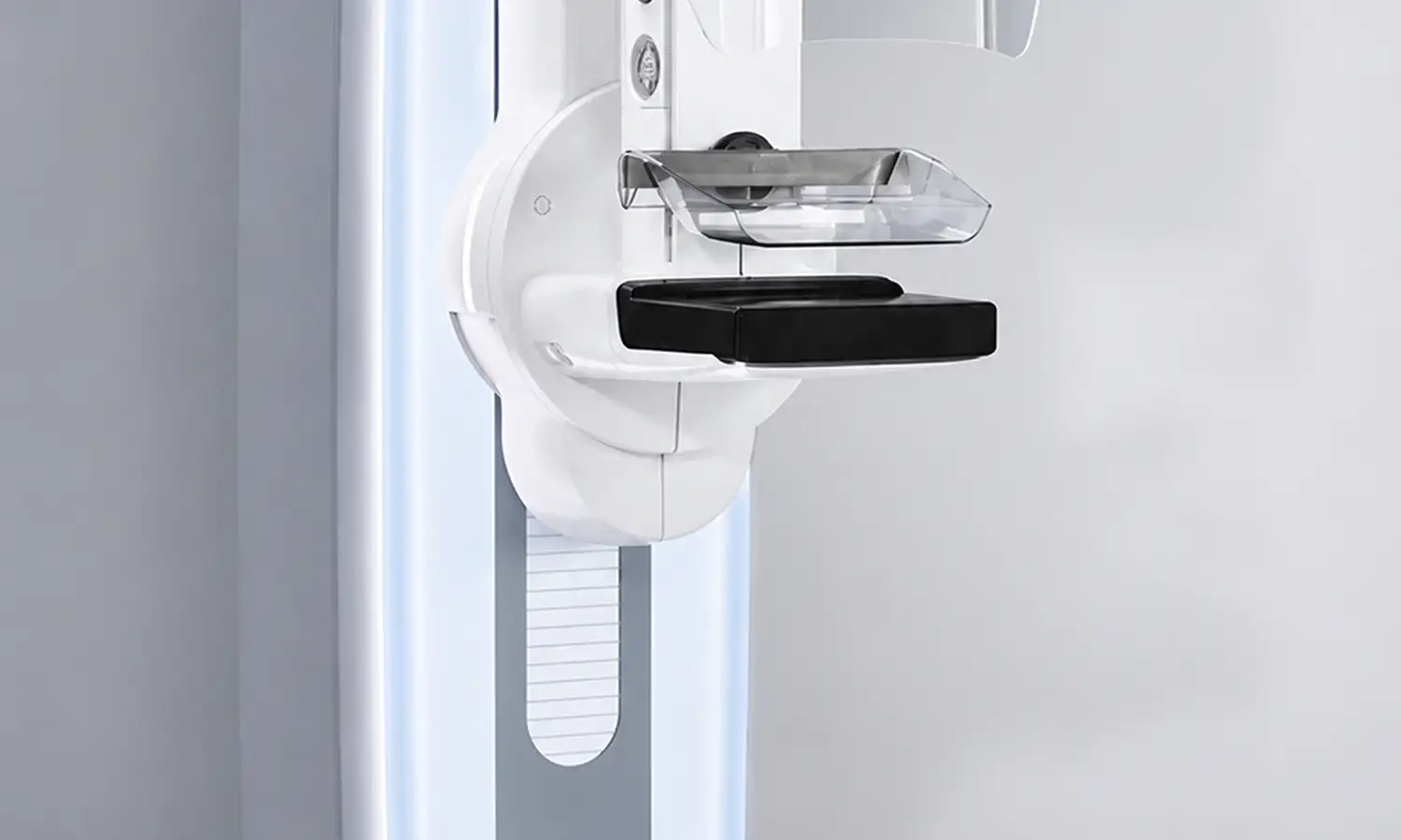
The MAMMOMAT system utilizes high-performance digital detectors and advanced image processing technology to capture high-resolution breast images, precisely visualizing tissue structures, microcalcifications, and potential abnormalities.Digital image processing algorithms optimize contrast and detail, allowing physicians to clearly identify imaging features associated with early-stage breast disease.During clinical examinations, the system adjusts imaging parameters to suit the specific breast tissue characteristics of each patient, ensuring high image quality while minimizing radiation exposure and enhancing the safety and reliability of the procedure.
2. 3D breast tomosynthesis technology
Selected models in the MAMMOMAT series support 3D breast tomosynthesis. By acquiring image data from multiple angles and performing 3D reconstruction, the system enables physicians to examine breast tissue structures across various planes.This technology effectively mitigates the effects of tissue overlap common in traditional 2D imaging and enhances the detection of abnormalities hidden within dense breast tissue—proving particularly valuable for assessing complex breast structures and identifying early-stage, minute lesions.The 3D tomosynthesis function provides physicians with richer imaging data, thereby increasing the accuracy of breast disease diagnosis.
3. Smart exposure control and low-dose imaging technology
Equipped with intelligent exposure control technology, the MAMMOMAT system automatically optimizes exposure parameters based on breast thickness, tissue density, and specific examination requirements, striking an optimal balance between image quality and radiation dose.By precisely controlling X-ray output and utilizing advanced image processing algorithms, the system minimizes unnecessary radiation exposure while meeting clinical diagnostic needs, thereby enhancing patient safety.Additionally, the system's ergonomic design improves comfort during the examination, helping to reduce patient anxiety and stress.
4. Adjunctive techniques for stereotactic biopsy
The MAMMOMAT system supports stereotactic biopsy capabilities, enabling physicians to precisely locate and sample abnormal areas identified in breast images.Through image-guided localization technology, physicians can accurately determine the spatial position of lesions, thereby enhancing the precision of needle biopsies.This technology is particularly valuable for assessing microcalcifications, early-stage abnormalities, and lesions that are difficult to access or palpate, facilitating a seamless workflow from initial imaging detection to precise diagnosis of breast diseases.
Scope of Indications
The Siemens MAMMOMAT digital mammography system is primarily used for the screening, diagnosis, and precise localization of breast diseases.
1. Areas for examination/adjunctive diagnosis
Field Primary Applications
Breast cancer screening Early breast cancer screening and regular check-ups for high-risk groups
Diagnosis of breast diseases Assessment of breast masses, abnormal calcifications, and structural changes
Preoperative assessment: determining the extent of the lesion prior to breast cancer surgery.
Biopsy localization: Precise localization and sampling of abnormal areas identified via imaging
2. Suitable patient population
• Individuals requiring early breast cancer screening
For asymptomatic individuals requiring breast cancer screening, the MAMMOMAT system utilizes high-resolution breast imaging to detect early-stage abnormalities, thereby improving the capability for early disease detection.
• Patients with abnormal breast imaging findings
For patients with abnormalities detected via breast ultrasound, clinical palpation, or prior examinations, this device provides highly detailed imaging information to assist physicians in determining the nature of the condition.
• Patients requiring precise biopsy localization
For patients with micro-calcifications or non-palpable abnormalities, the stereotactic capabilities of the MAMMOMAT system assist physicians in precisely locating lesions, thereby supporting subsequent pathological diagnosis.
Core Strengths
Comparison Dimension Traditional Breast Examination Methods Siemens MAMMOMAT Digital Mammography System
Imaging mode Primarily 2D imaging Supports both 2D and 3D tomographic imaging
Lesion visualization Susceptible to tissue overlap Multi-slice imaging improves visualization capabilities
Image quality depends on equipment performance; features high-definition digital image processing.
Radiation control Limited parameter adjustment Intelligent exposure optimization and low-dose control
Biopsy localization: Limited localization accuracy vs. Support for precise stereotactic localization
Clinical Application: Standalone examination; integrated application for screening, diagnosis, and localization
Conventional Breast Examination Methods
Conventional breast examinations rely mainly on two-dimensional imaging for assessment, where the overlapping of breast tissue can obscure the visualization of certain lesions.In complex cases or when screening for early-stage, minute lesions, the limited dimensionality of the imaging data presents challenges in accurately determining the extent and location of the lesions.Furthermore, the adjustment of parameters and the localization for biopsies typically rely on conventional imaging guidance, which imposes certain limitations on precision.
Siemens MAMMOMAT Digital Mammography System
The Siemens MAMMOMAT digital mammography system utilizes high-definition digital imaging technology to enhance the visualization of breast tissue and abnormal lesions. It supports both 2D imaging and 3D tomosynthesis modes, providing richer imaging data for clinical screening, diagnosis, and treatment decisions.Compared to traditional breast imaging methods, this system uses multi-layer imaging to reduce the impact of tissue overlap and improve the detection of tiny lesions.Additionally, intelligent exposure optimization technology adjusts imaging parameters based on examination requirements, optimizing radiation control while maintaining image quality.The system also supports stereotactic-guided biopsy, enhancing lesion localization accuracy and facilitating the early detection and precise treatment of breast diseases.
FAQ
1. What is digital mammography primarily used for?
Mammography can be used for breast cancer screening and to help evaluate breast lumps, nipple discharge, localized pain, or other abnormalities.Physicians determine whether to combine this with ultrasound, MRI, or other examinations based on factors such as age, symptoms, breast density, and previous test results.
2. Why is breast compression necessary during the examination?
Appropriate compression spreads out the breast tissue, reduces tissue overlap and motion blur, and helps control radiation dosage while maintaining image quality. The compression duration is usually brief; if significant pain occurs, please communicate immediately with the operator.
3. Does a mammogram involve radiation?
The procedure utilizes low-dose X-rays, involving a small amount of ionizing radiation.Equipment settings and operational protocols are designed to minimize radiation exposure while ensuring diagnostic requirements are met; pregnant women or those who may be pregnant should inform the medical staff beforehand so the physician can weigh the necessity of the examination.
4. Does the menstrual cycle affect the examination experience?
Some women experience breast tenderness or swelling before menstruation, making compression during examinations potentially more uncomfortable.If the examination is not urgent, you may inquire about scheduling it for a time when breast sensitivity is lower; however, do not delay seeking medical attention if suspicious symptoms are detected.
5. How do I schedule a Siemens MAMMOMAT mammography exam?
Before making an appointment, please inform the breast specialist or radiology department whether the visit is for routine screening or due to symptoms such as a lump or nipple discharge, and disclose any pregnancy or potential pregnancy.On the day of the examination, please bring previous breast imaging records and reports; avoid using talcum powder, antiperspirants, or skincare products containing metal particles on the underarms or around the breasts.